From a medical point of view, computerized tomography is nowadays a commonly available and very sophisticated examination method that, using X-rays and mathematical analysis, can display the internal structure of the scanned object. Although CT scanning is associated with science and technology, it would surprise many that its humble beginnings could be rooted in rock and roll.
The inventor of the CT scanner is Sir Godfrey Hounsfield. The youngest son of Thomas Hounsfield's five children, he was born in August 1919. In the farm environment of Nottinghamshire in central England, little Godfrey found many opportunities for various adventurous expeditions and ingenious explorations. He built electric recording devices, built a projector for the village cinema, shot barrels filled with water up to 300 meters high with an acetylene explosion. Despite his obvious aptitude for physics and mathematics, Godfrey was not one of the top students at Magnus High School. On the outbreak of the Second World War he joined the Royal Air Force as a radar instructor and moved via the Royal College of Science to the Cranwell Radar School.
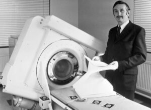
In 1949, Godfrey Hounsfield joined Electrical and Musical Industries (EMI), based in Hayes, on the western outskirts of London. EMI's roots date back to the end of the 19th century, when the gramophone method of recording and reproducing sound using discs was invented. EMI itself was created during the Great Depression in 1931 by the merger of two companies (The Gramophone Company and The Columbia Graphophone Company). In the 20th century, the music industry experienced century boom in the 50s and 60. years, but let's go back to Sir Hounsfield, because it was EMI that was getting rich at that time from the sale of The Beatles' recordings. The expensive research, sponsored by EMI, was born in Hounsfield's head during a weekend walk in the countryside, just as the Beatles were preparing the famous album 'Sgt. Pepper's Lonely Hearts Club Band' - in June 1967. Reflecting on his research into radar systems emitting radio waves from a central point in all directions and looking for structures around them, Hounsfield thought, "Why not try the reverse approach—broadcasting from the outside and detecting hidden structures inside?" He decided to examine three-dimensional objects as a series of two-dimensional sections using X-rays, during which he will measure the attenuation of the radiation in all directions of its passage and then reconstruct the image of each section and the three-dimensional image of the object in a computer. As an employee of an industrial company and essentially a reclusive inventor, Hounsfield remained distant from the research activity and working relationships of other researchers in the field. He did not even have a general overview of the relevant literature. He did not know the article by Děčín native and Viennese mathematician Johann Radon from 1917 on the reconstruction of the image of a three-dimensional object from an infinite number of plane projections. He did not know the Los Angeles neurologist William Oldendorf's publication from 1961 presenting a prototype of a simple tomograph in which the radiation source and detector rotate around the object being examined. He was also unaware of American physicist Allan Macleod Cormack's work in 1963 and 1964, which showed that changes in tissue density could be calculated from X-ray data, but due to limitations in computing power, he was not in the 60s. years, no machine has been constructed. Independently of everyone, with the use of gamma radiation and computers at the then early stage of development, he started research in 1967, for which he procured a lathe bed and a source of concentrated gamma radiation (americium 241) to rotate around the scanned objects (bottles, pieces of Plexiglas, later a frontal cut brain removed from the skull). The result of approximately 28 measurements in 000 days and 9 hours of computer reconstruction at a slice thickness of 2,5 mm, an acquisition time of over 13 minutes, a reconstruction time of 4 seconds, and a resolution of 20 × 80 display point-pixels of 80 × 3 mm was an image with sufficient contrast to distinguishing between gray and white brain matter. Hounsfield later replaced the gamma radiation source with an X-ray and achieved a reduction in measurement time to 9 hours. This invention-method of transverse axial tomography was patented in 1968. In the early seventies of the 20 century, the vast majority of the then highly conservative radiological community clung to the old X-ray images and were unable to appreciate the contribution of Hounsfield's invention. His images did not arouse much interest even at the European Radiological Congress in Amsterdam in the summer of 1971.
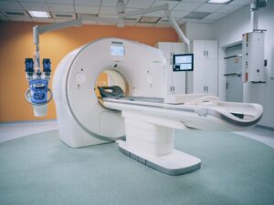 On October 1, 1971, at Wimbledon's Atkinson Morley Hospital, he performed the first clinical examination of a human using computed tomography, for which neuroradiologist James Ambrose selected a woman with a suspected tumor in the frontal lobe of the brain. The CT examination clearly showed the cyst, and also in the following dozen patients, the method reliably revealed brain diseases in various forms. At the end of 1973, the first commercial EMI CT 1000 scanner was launched, with the acquisition time reduced to 20 and the number of detectors increased to 30, which enabled image reconstruction with a resolution of 320 × 320 pixels (current CT systems can, under specific conditions, perform a full-body scan in 1 second, using a set of up to 320 detectors and a resolution of 1048 x 1048 pixels).
In 1979, when around 1000 sensors were already working in the world, the Nobel Prize in Physiology or Medicine was awarded "for the development of computed tomography." The laureates became, as expected, Hounsfield and, for many, unexpectedly, Cormack. On December 8, Hounsfield gave the Nobel lecture (Computed Medical Imaging. Med Phys 1980; 7: 283–290, Science 1982; 210: 22–28) and met a second laureate for the first time in his life. A shy Hounsfield asked Cormack to deliver the three-minute Nobel acceptance speech for both of them. Cormack complied, and at the banquet he again spoke for both of them: "It is no exaggeration to say that what Hounsfield and I know of medicine and physiology could fit on a small recipe book." After receiving the Nobel Prize, Housnfield's advice to the young was: " Don't worry if you can't pass the exams if you feel you have understood the topic”.
Finally, it is generally believed that the proceeds from EMI's sales of The Beatles' records enabled the company to fund its Central Research Laboratory (CRL) and participate in the development of the computed tomograph. Some have defined it as the Beatles' gift to medicine. In the past, however, analysis has shown that these claims are false. Excluding the labor costs of Godfrey Hounsfield and his team, the EMI CT scanner cost around £100 to develop. The cost to the UK DHSS (Department Of Health and Social Security) was £000. Thus, the financial contribution of DHSS to the development of the CT scanner was significantly greater than that of EMI. The positive aspect of this misconception is that it preserves the name of the company that developed the CT scanner in public memory.