Department of Nuclear Medicine and Endocrinology, 2nd Medical Faculty, Charles University and University Hospital Motol
Our department focuses on the diagnosis, treatment and dispensary of patients with thyroid tumors that can be treated with nuclear medicine and on patients with more severe forms of thyroid orbitopathy. In the field of nuclear medical diagnostics, we provide services in a wide range of the entire field, in the field of endocrinology, consulting services within the hospital and superconsulting service for catchment endocrinologists specialized in thyrology.
prof. MUDr. Petr Vlcek, CSc., MHA
(224) 434 600
petr.vlcek@fnmotol.c
Primary
MD Kateřina Táborská
(224) 434 626
katerina.taborska@fnmotol.cz
Head nurse
Ladislava Novotná
(224) 434 605
ladislava.novotna@fnmotol.cz
Secretariat
224 434 601, 224 434 602
fax: +224 434 620 XNUMX
nuclear@fnmotol.cz
Where to find us:
Endocrinology outpatient clinic: adult part, node A, 3rd floor
Nuclear medicine ambulance: adult section, node B, -2. floor
Bed department: adult section, node B, -1. and -2. floor
Clinic management: adult part, node B, -2. floor
The NM ambulance is located in communication node B, -2. floor. It provides outpatient examinations for the University Hospital in Motol and the catchment area, offers a wide range of scintigraphic examinations (skeletal scintigraphy, nuclear nephrology, cardiology, tumor diagnostics, pneumology, CNS examinations, thyroid diagnostics) in patients of all ages, as well as palliative treatment of bone metastases and radionuclide synovectomy. The NM ambulance cooperates with surgical workplaces during radiation-guided procedures.
All examinations are performed on the recommendation of the attending physician after prior order.
Operation is provided by doctors (certified from NM), nurses for NM, radiology assistants. Radiopharmaceuticals are prepared in the radiopharmaceutical laboratory, which is part of the NM outpatient clinic. The Department of Radiological Physics KNME cooperates in providing diagnostic and treatment procedures using ionizing radiation sources.
KNME primary care physician and KNME nuclear medicine outpatient physician:
MD Kateřina Táborská | (224) 434 626 |
Doctors:
| MD Lucie Lančová | (224) 434 626 | lucie.lancova@fnmotol.cz |
MD Katerina Michalová | (224) 434 626 | |
MD Jitka Svobodová | (224) 434 626 | |
MD Zuzana Hotváthová | (224) 434 626 |
Office hours
Ordering scintigraphic examinations based on the recommendation of the attending physician by telephone Mon-Thu: 8:00 - 16:00, Fri 8:00-14:00 on telephone number (224) 434 626.
| Monday to Thursday | 7:00 - 17:30 |
| Friday | 7:00 - 14:00 |
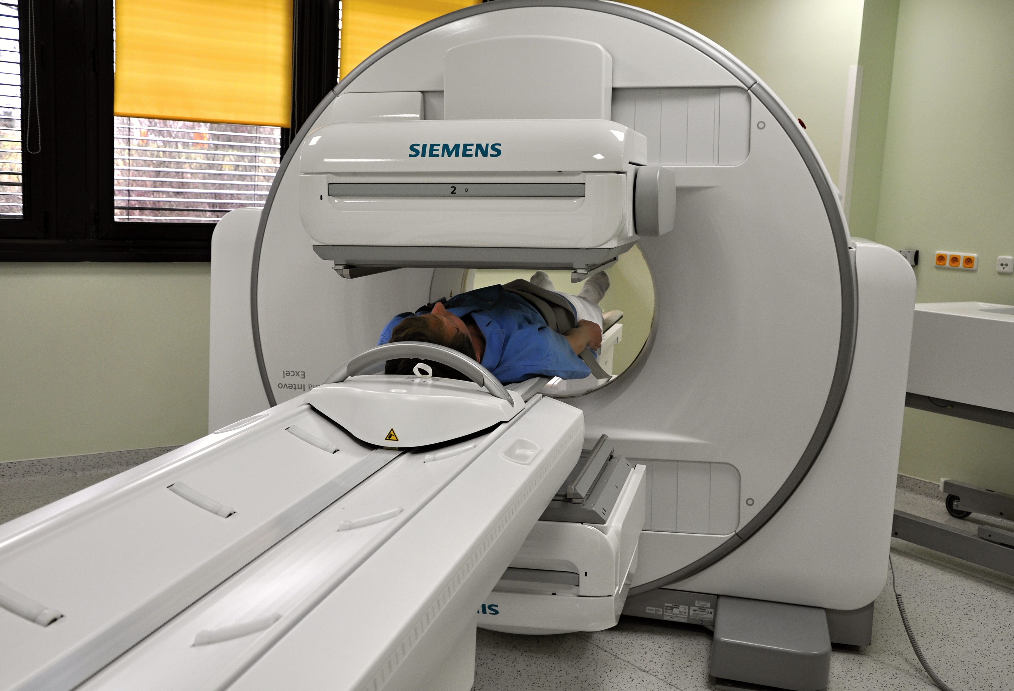
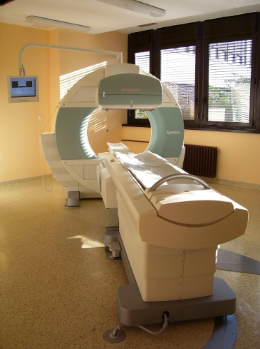
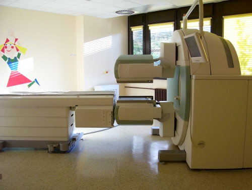
Instrumentation
Scintigraphic examinations are performed on the following devices:
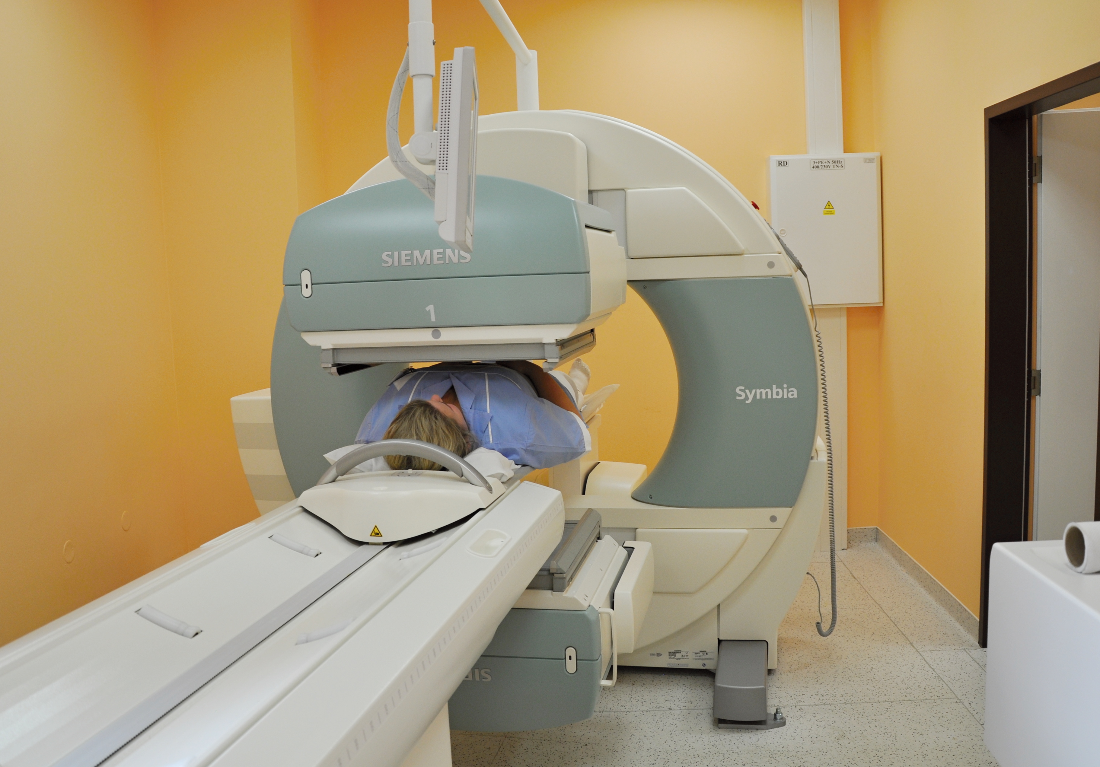
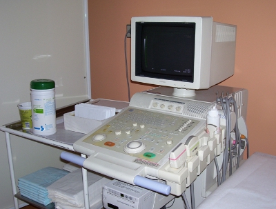
| Symbia Intevo Excel hybrid SPECT / CT camera | SYMBIA S double head gamma camera | SYMBIA T hybrid SPECT / CT camera |
 |  |  |
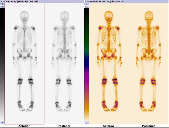
1. Skeletal scintigraphy
Your doctor has ordered a scintigraphic examination to examine your metabolic bone activity, which may identify possible bone problems related to your condition.
The examination is associated with radiation exposure, which is comparable to other X-ray examinations and which you will receive in two years of your life from cosmic radiation and radiation from natural radioactive sources.
Preparation: Not necessary, you can eat and drink before the examination. After administration of the radioactive substance, drink more (at least 0,5 l of fluid) and urinate frequently, observe this precaution during the day after the examination.
Design: You will receive a small amount of radioactive substance in an intravenous injection. The test itself is performed 3-5 hours after the injection and lasts approximately 30-90 minutes.
2. Targeted three-phase scintigraphy of the skeleton
Your doctor has ordered a scintigraphic examination to examine your metabolic bone activity and to identify your possible bone problems.
The examination is associated with radiation exposure, which is comparable to other X-ray examinations and which you will receive in two years of your life from cosmic radiation and radiation from natural radioactive sources.
Preparation: Not necessary, you can eat and drink before the examination. After administration of the radioactive substance, drink more (at least 0,5 l of fluid) and urinate frequently, observe this precaution during the day after the examination.
Design: You will receive a small amount of radioactive substance in an intravenous injection directly under the camera. In this first phase, the blood supply to the examined area is monitored. The next part of the examination is performed 3-5 hours after the injection and lasts 30-90 minutes.
3. Bone marrow scintigraphy
Your doctor has ordered a scintigraphic examination to show your bone marrow.
The examination is associated with radiation exposure, which is comparable to other X-ray examinations and which you will receive in two years of your life from cosmic radiation and radiation from natural radioactive sources.
Preparation: Not necessary
Design: You will receive a small amount of radioactive substance in an intravenous injection. The examination itself is performed 1-2 hours after the injection and lasts 30-90 minutes.
1. Perfusion scintigraphy of the lungs
Your doctor has ordered a scintigraphic examination to monitor your blood flow to your lungs.
The examination is associated with radiation exposure, which is comparable to other X-ray examinations and which you will receive in two years of your life from cosmic radiation and radiation from natural radioactive sources.
Preparation: Not necessary
Design: You will receive a small amount of radioactive substance in an intravenous injection. The test itself is performed immediately after the injection and lasts approximately 15-30 minutes.
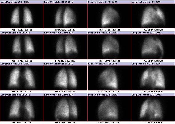
2. Ventilation scintigraphy of the lungs
Your doctor has ordered a scintigraphic examination to monitor your airflow to your lungs.
The examination is associated with radiation exposure, which is comparable to other X-ray examinations and which you will receive in two years of your life from cosmic radiation and radiation from natural radioactive sources.
Preparation: Not necessary. In patients with chronic obstructive pulmonary disease, administration of bronchodilators half an hour before the examination is appropriate.
Design: You will inhale a small amount of radioactive material for 10 minutes. Inhalations and examinations take place either simultaneously or follow each other. The examination itself then takes about half an hour.
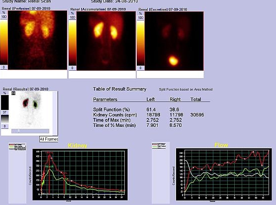
1. Dynamic scintigraphy of the kidneys
Your doctor has ordered a scintigraphic examination to monitor your kidney function and urine outflow through the hollow kidneys.
The examination is associated with minimal radiation exposure. In well-functioning kidneys, 95% of the administered amount is excreted within 4 hours after administration.
Preparation: You can eat and drink, on the contrary, it is recommended to be well watered when you arrive for the examination. In the morning on the day of the examination, drink enough (at least two large glasses of water, juice) and after arriving at the ward you will drink another 0,5 liters of fluid.
In young children, fluid intake is proportional to their age and weight (100 ml fluids / 10 kg body weight), children are breastfed in the usual way and it is advisable to breastfeed the child upon arrival at the examination.
Design: You will receive a small amount of radioactive substance in an intravenous injection directly on the examination table. The examination itself lasts 20 minutes, after which a picture is taken after urination. If a blockage of urine is suspected, you may be given a substance that accelerates the outflow of urine (diuretic) and the injection is followed by additional pictures lasting 20 minutes.
2. Static scintigraphy of the kidneys
Your doctor has ordered a scintigraphic examination to monitor your kidney function.
The examination is associated with radiation exposure, which is comparable to other X-ray examinations and which you will receive in two years of your life from cosmic radiation and radiation from natural radioactive sources.
Preparation: Not necessary. You can eat and drink. After administration of the radioactive substance, drink more (at least 0,5 l of fluids) and urinate frequently, follow this precaution during the day after the examination.
Design: You will receive a small amount of radioactive substance in an intravenous injection. The test itself is performed 2,5-4 hours after the injection and lasts approximately 30-90 minutes.
3. Glomerular filtration of 99mTc DTPA
Your doctor has ordered a scintigraphic examination to determine your kidney function by taking two blood samples.
The examination is associated with minimal radiation exposure. In well-functioning kidneys, 95% of the administered amount is excreted within 4 hours after administration.
Preparation: Not necessary. You can eat and drink.
Design: You will receive a small amount of radioactive substance in an intravenous injection. Blood samples are taken 120 and 180 minutes after administration.
4. Functional scintigraphy of the transplanted kidney
Your doctor has ordered a scintigraphic examination to monitor the blood supply and function of the transplanted kidney.
The examination is associated with radiation exposure, which is comparable to other X-ray examinations and which you will receive in two years of your life from cosmic radiation and radiation from natural radioactive sources.
Preparation: You can eat and drink, on the contrary, it is recommended to be well watered when you arrive for the examination. In the morning on the day of the examination, drink enough (at least two large glasses of water, juice) and after arriving at the ward you will drink another 0,5 liters of fluid. Applies to adult patients unless the treating physician has restricted fluid intake.
In young children, fluid intake is proportional to their age and weight (100 ml fluids / 10 kg body weight).
Design: You will receive a small amount of radioactive substance in an intravenous injection directly on the examination table. The examination itself lasts 20 minutes, after which a picture is taken after urination.
5. Direct radionuclide cystography
Your doctor has ordered a scintigraphic examination to monitor your urinary tract (bladder and ureters).
The examination is associated with a minimum radiation exposure, which is lower compared to X-ray examination.
Preparation: Insertion of a urinary catheter in the department that orders the examination.
Design: While lying on the examination table, you will receive a small amount of radioactive substance through a catheter inserted into the bladder. After the bladder is sufficiently full, urination is recorded sitting or standing.
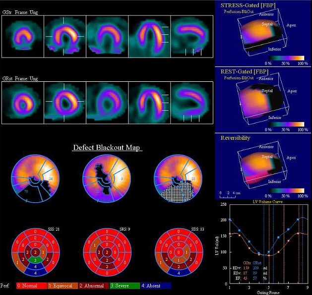
1. Perfusion myocardial SPECT after exercise
Your doctor has ordered a scintigraphic examination, which examines the blood supply to the heart muscle during exercise (ergometric = pedaling) and can also provide information about heart function.
The examination is associated with radiation exposure, which is comparable to other X-ray examinations and which you will receive in two years of your life from cosmic radiation and radiation from natural radioactive sources.
Preparation: Do not eat, drink (drink only mineral water), do not smoke for 2 hours before the examination.
! Not valid for diabetics who follow their regimen!
Discontinue:
- nitrates 24 hours
- beta-blockers at least 2 days in advance, in hypertensive patients replace the beta-blocker with another antihypertensive agent
If your doctor wants to check your blood flow to the myocardium during medication, do not stop taking beta-blockers and nitrates.
Do not miss other medicines - take them on the day of the examination!
Bring comfortable sports shoes, snacks, including drinks, medication
Design: You will receive a small amount of radioactive substance in an intravenous injection during an ergometric load. The examination itself is performed 15-60 minutes after the end of physical activity and lasts approximately 30 minutes. Total examination time 3-5 hours. In some cases, it is necessary to supplement the rest examination, which is performed on another day, with a minimum interval of two days from the load.
2. Perfusion myocardial SPECT after dipyridamole exposure
Your doctor has ordered a scintigraphic examination to check the blood supply to the heart muscle during exercise (pharmacological = administration of a substance that causes increased blood flow to the heart muscle) and may also provide information on heart function.
The examination is associated with radiation exposure, which is comparable to other X-ray examinations and which you will receive in two years of your life from cosmic radiation and radiation from natural radioactive sources.
Preparation: It is necessary to omit some drugs and foods that may affect the examination!
1 day must omit coffee, tea, chocolate, cocoa, caffeinated beverages (coca-cola, energy drinks), bananas. Medication is prescribed by your doctor.
- drugs containing methylxanthines for at least 2 days (eg Afonilum, Euphyllin, Spophyllin, Teotard, Theoplus, Aminophyllin, Syntophyllin…), dipyridamole in oral form for 24 hours
- caffeine drugs (analgesics eg Acifein, Alnagon, Ataralgin, Coldrex, Ibufein, Panadol extra ..., Kinedryl)
- medicines with pseudoephedrine (eg Clarinase, Disophrol, Modafen, Nurofen stopgrip, Panadol plus grip, Paralen plus ...)
- no need to discontinue pentoxifylline (eg Agapurin, Trental, Pentomer) and clopidogrel (Plavix)
- adjustment of cardiac medication - nitrates discontinued 24 hours. Beta-blockers do not need to be discontinued.
- fasting at least 2 hours before exercise, drink only mineral water
- children last meal 2 hours before the examination, they have to drink
Bring a snack, including a drink.
Design: The test is started by intravenous administration of dipyridamole for 4 minutes. In another 3-4 minutes you will receive a small amount of radioactive substance in an intravenous injection. The examination itself is performed 30-60 minutes after the end of the pharmacological load and lasts approximately 30 minutes. Total examination time 3-5 hours. In some cases, it is necessary to supplement the rest examination, which is performed on another day, with a minimum interval of two days from the load.
3. Perfusion myocardial SPECT after dobutamine exposure
Your doctor has ordered a scintigraphic examination to check the blood supply to the heart muscle during exercise (pharmacological = administration of a substance that causes increased cardiac work) and may also provide information on heart function.
The examination is associated with radiation exposure, which is comparable to other X-ray examinations and which you will receive in two years of your life from cosmic radiation and radiation from natural radioactive sources.
Preparation: Do not eat, drink (drink only mineral water), do not smoke for 2 hours before the examination.
! Not valid for diabetics who follow their regimen!
It is necessary to omit some drugs that may affect the examination. Medications are stopped by your doctor.
- nitrates 24 hours
- beta-blockers at least 2 days in advance, in hypertensive patients replace the beta-blocker with another antihypertensive agent
Bring a snack, including drinks and medication.
Design: The examination is started by intravenous administration of dobutamine for 6-12 minutes. You will receive a small amount of radioactive substance intravenously before the end of the infusion. The examination itself is performed 30-60 minutes after the end of the pharmacological load and lasts approximately 30 minutes. Total examination time 3-5 hours. In some cases, it is necessary to supplement the rest examination, which is performed on another day, with a minimum interval of two days from the load.
4. Perfusion myocardial SPECT at rest
Your doctor has ordered a scintigraphic examination, which examines the blood supply to the heart muscle at rest and can also provide information about heart function.
The examination is associated with radiation exposure, which is comparable to other X-ray examinations and which you will receive in two years of your life from cosmic radiation and radiation from natural radioactive sources.
Preparation: Do not eat, drink (drink only mineral water), do not smoke for 2 hours before the examination!
! Not valid for diabetics who follow their regimen!
The drugs are not discontinued. Bring a small snack, yogurt or milk.
Design: You will receive a small amount of radioactive substance in an intravenous injection. The examination itself is performed in 60-90 minutes and lasts approximately 30 minutes.
5. First pass angiocardiography (first pass)
Your doctor has ordered a scintigraphic examination to monitor the first flow of radioactive material through the heart and lungs, to assess the function of the left and right ventricles and to assess the presence of short-circuit defects.
The examination is associated with radiation exposure, which is comparable to other X-ray examinations and which you will receive in two years of your life from cosmic radiation and radiation from natural radioactive sources.
Preparation: Thyroid blockade by administration of Chlorigen (performed after arrival for examination).
Design: You will receive a small amount of radioactive substance in an intravenous injection. The test itself is performed immediately after the injection and lasts 1 minute.
6. Radionuclide equilibrium ventriculography
Your doctor has ordered an examination to assess the function of your heart chambers.
The examination is associated with radiation exposure, which is comparable to other X-ray examinations and which you will receive in two years of your life from cosmic radiation and radiation from natural radioactive sources.
Preparation: Thyroid blockade by administration of Chlorigen (performed after arrival for examination).
Design: We will mark your red blood cells directly in your bloodstream by giving two intravenous injections 30 minutes apart. The examination itself takes approximately 30 minutes.
1. Lymphoscintigraphy of the sentinel node
Your doctor has ordered a scintigraphic examination to show a sentinel node that has a direct inflow of lymph from the tumor.
The examination is associated with radiation exposure, which is significantly lower compared to other X-ray examinations.
Preparation: Not necessary.
Design: The day before or on the day of the operation, you will be given a small amount of radioactive substance in the vicinity of the tumor, which spreads from the injection site through the lymphatic vessels to the guard node. Depending on the type of tumor, the scan begins immediately or up to 2 hours after administration, a scan is taken lasting 3 minutes and the doctor marks the node on the skin, sometimes it is necessary to repeat the images until the node is imaged. The operating doctor uses a special hand probe to find the node during the operation. Based on the histological examination, the further procedure of the operation and treatment can be determined.
2. Tumor scintigraphy - 99mTc MIBI
Your doctor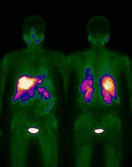 I ordered a scintigraphic examination, in which a substance called MIBI is administered, which has the ability to accumulate in some tumor tissues.
The examination is associated with radiation exposure, which is comparable to other X-ray examinations and which you will receive in two years of your life from cosmic radiation and radiation from natural radioactive sources.
Preparation: Not required.
Execution: You will receive a small amount of radioactive substance in an intravenous injection, which is usually given into a vein in the back of the foot. The test itself is performed 10 minutes after the injection and lasts 30-90 minutes.
3. Tumor scintigraphy - 123I MIBG
Your doctor has ordered a scintigraphic test to give you a substance called MIBG, which has the ability to accumulate in neuroendocrine tumors.
The examination is associated with radiation exposure, which is comparable to other X-ray examinations and which you receive in a year of your life from cosmic radiation and radiation comes from natural radioactive sources.
Preparation: You can eat and drink before you arrive. Thyroid blockade: starts the day before application and lasts for 3 days. If the patient is after a total thyroidectomy, the blockade is not performed. Chlorigen or pharmacy-prepared perchlorate can be obtained from a nuclear medicine clinic.
Chlorogen:
| grown-ups | 400 mg / day (4 capsules / day) |
| children 1-6 years | 100 mg / day (1 capsules / day) |
| children 6-15 years | 200 mg / day (2 capsules / day) |
Lugol's solution: 1 drop / 3 kg body weight
Discontinuation of drugs affecting MIBG uptake (performed by the attending physician):
If you are taking medication, check it with your doctor at least 4 weeks before the examination and stop it according to his recommendation! Discontinued antihypertensives can be replaced by alpha-blockers (Cardura, Zoxon), diuretics.
- Combined alpha / beta blockers: labetalol 72 hours, others 24 hours
- Calcium channel blockers: 24-48 hours
- sympathomimetics: phenylpropanolamine, pseudoephedrine, phenyllephrine, amphetamine, dopamine, Fenoterol (Berotec®), Salbutamol (Ventolin®), Terbutaline (Bricanyl®) nasal drops and sprays with Xylometazoline (Otrivine drops®). 24 hours, for the treatment of glaucoma Brimonidine 48 hours
- antipsychotics (neuroleptics): Levomepromazine, Amisulpride 72 hours, Clozapine 7 days, Olanzapine 7-10 days, risperidone 5 days, depot form 1 month, sertindole 15 days, zotepine 5 days, Haloperidol, Flupentixol, Fluphenazine 2 days, depot form 1 month , Chlorpromazine, prochlorperazine 24 hours
- tricyclic and other antidepressants: amitriptyline and derivatives, imipramine and derivatives 24-48 hours, mirtazapine 8 days, maprotiline, mianserin, trazolone, venlaflaxine 48 hours
- opioids: tramadol 24 hours, CNS stimulants: Atomexetine 5 days, Modafinil 72 hours, methylphenidine 48 hours
Increased fluid intake is recommended after application. If you do not suffer from diarrhea, take the medicine to empty your bowels (glycerin suppository, Guttalax) in the evening after application.
Design: The examination is performed in two days. The first day you will receive a small amount of radioactive substance by intravenous injection. The examination itself is performed 24 hours apart and lasts 30 minutes to 2 hours, depending on the number of images.
4. -111In OctreoScan tumor scintigraphy
Your doctor has ordered a scintigraphic test to give you a substance called Octreoscan, which has the ability to accumulate in neuroendocrine tumors.
The test is associated with a radiation exposure that is higher than other X-ray tests and is approximately seven times the dose you receive in a year of your life from cosmic radiation and radiation from natural radioactive sources.
Preparation: Omission of somatostatin preparations (Sandostatin 3 days, Somatulin 6 weeks) will be ensured by the sending doctor. You can eat and drink before the test. After administration of the radioactive substance, drink more (at least 0,5 l of fluid) and urinate frequently, observe this precaution during the day after the examination.
Design: You will receive a small amount of radioactive substance in an intravenous injection. The examination itself is performed in 2-3 days, the first images are performed 4-6 hours after application, the next in 24, sometimes 48 hours, and each scan lasts 1-2 hours.
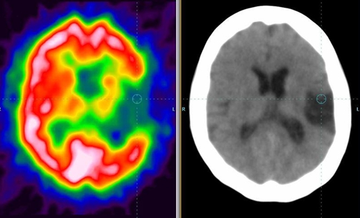
1. Perfusion SPECT brain
Your doctor has ordered a scintigraphic examination of your brain, which assesses the blood supply and functional condition of the cerebral cortex.
The examination is associated with radiation exposure, which is comparable to other X-ray examinations and which you will receive in two years of your life from cosmic radiation and radiation from natural radioactive sources.
Preparation: Before coming for the examination
Skip alcohol and caffeine preparations (coffee, coca-cola, energy drinks) 24 hours before the examination.
Procedure: You will receive a small amount of radioactive substance in an intravenous injection. The injection is given at rest, so we will insert a cannula, place you in a darkened room where you will rest, and you will not talk for about 10-15 minutes before and after the injection. The examination itself starts 45-60 minutes after application and lasts approximately 45 minutes, after which you will lie on the bed, the camera will move around your head. It is important that you stay still during the examination.
2. Perfusion SPECT of the brain after Diamox load
Your doctor has ordered a scintigraphic examination of your brain after administration of Diamox. This substance increases the blood supply to your brain, there is no increase in the areas behind the narrowing of the arteries.
The examination is associated with radiation exposure, which is comparable to other X-ray examinations and which you will receive in two years of your life from cosmic rays.
Preparation: Before arriving for the examination, omit alcohol and caffeine preparations (coffee, coca-cola, energy drinks) 24 hours before the examination.
Design: The examination is performed in 2 days, the first resting examination, when you receive a small amount of radioactive substance in an intravenous injection. The injection is given at rest, so we will insert a cannula, place you in a darkened room where you will rest and will not talk for about 10-15 minutes before and after the injection. The examination itself starts 45-60 minutes after application and lasts 45 minutes, during which you will lie on the bed, the camera will move around your head. It is important that you stay still during the examination. The next day we will perform a stress test, when you will first receive Diamox intravenously, we will check your blood pressure regularly during the administration and you will receive an injection with a radioactive substance in 20 minutes as described above.
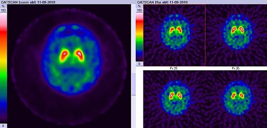
3. 123I - DaTSCAN
Your doctor has ordered a scintigraphic examination to assess the biochemical and structural integrity of the presynaptic dopaminergic system.
The examination is associated with radiation exposure, which is comparable to other X-ray examinations and which you receive in two years of your life from cosmic radiation and radiation comes from natural radioactive sources.
Preparation: omit drugs that affect the binding of dopamine transporters before the examination (performed by the attending physician):
- amphetamine, cocaine
- benzotropine (Apo-benzotropine)
- bupropion (Wellbutrin, Zyban)
- Mazindol
- methylphenidine (Ritalin)
- sertraline (Adjuvin, Apo-Sertral, Asentra, Sertraline, Serlift, Zoloft)
Before and after RF application:
- thyroid blockade: Chlorigen (Perchlorate) 30 minutes before (400mg) and 12-24 hours after radiopharmaceutical application (400mg) - performed after arrival for examination
After administration drink more radioactive substances (at least 0,5 l of fluid) and urinate frequently, observe this precaution during the day after the examination.
Design: You will receive a small amount of radioactive substance in an intravenous injection. The examination itself is performed 3 to 6 hours after application and lasts 45 minutes.
Scintigraphy of inflammation with labeled leukocytes
Your doctor has ordered a scintigraphic examination to check that you do not have a site of inflammation in your body in which white blood cells (leukocytes) accumulate.
The examination is associated with radiation exposure, which is comparable to other X-ray examinations and which you receive in two years of your life from cosmic radiation and radiation comes from natural radioactive sources.
Preparation: Do not eat 4-6 hours before taking blood, you can drink. The sending doctor will arrange the KO + diff. Examination, bring the results (not older than 10 days) with you.
Design: According to the method of marking white blood cells, you will either receive a small amount of radioactive substance in an intravenous injection and the blood cells will be marked directly in your body or we will take 45-60 ml of blood, from which the white blood cells will separate, label with a radioactive substance. We will return the marked blood cells to you by intravenous injection.
The examination itself is performed every two days, scanning is performed repeatedly 1, 5-8 and 24 hours after the injection. Individual images can take from 10 minutes to 2 hours. In some cases, a bone marrow scintigraphic examination is completed at least two days later if an endoprosthesis infection is suspected.
1. Thyroid scintigraphy 99mTc pertechnetate
Your doctor has ordered a thyroid scintigraphy.
The examination is associated with radiation exposure, which is comparable to other X-ray examinations and which you receive in four years of your life from cosmic radiation and radiation from natural radioactive sources.
Preparation: It is necessary to discontinue thyroid medication before the examination (provided by the indicating doctor):
- Levotyroxine (Euthyrox, Letrox, Thyroid) for at least 10 days
- Triiodothyronine at least 3 days
Avoid administration of iodine in food, drugs for internal or external use at least 4 weeks before the examination (therapy with amiodarone, betadine, administration of X-ray contrast agent, application of iodine disinfectants on the skin may affect the accumulation of RF for up to 3 months).
You can eat, you can drink.
Design: You will receive a small amount of radioactive substance in an intravenous injection. The test itself is performed 10 minutes after the injection and lasts 20 minutes.
2. Thyroid scintigraphy 123 I
Your doctor has ordered a thyroid scintigraphy.
The examination is associated with radiation exposure, which is comparable to other X-ray examinations and which you receive in four years of your life from cosmic radiation and radiation from natural radioactive sources.
Preparation: It is necessary to stop thyroid medication before the examination (provided by the indicating doctor):
- Thyroxine (Euthyrox, Letrox, Thyroid) for at least 4 weeks
- Triiodothyronine for at least 17 days
- Propylthiouracil (Propycil), methimazole (Thyrozole, Favistan), Carbimazole at least 3 days - (examination in 72h rebound thyrostatics).
Exclude iodine administration in any application form at least 4 weeks before the examination.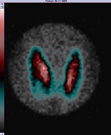
With a higher amount of iodine (therapy with amiodarone, betadine, KI, Solutan, administration of X-ray and CT iodine contrast agent, chronic application of iodine disinfectants on the skin, Jox spray, etc.) delay at least 3 months!
Fast for 4 hours before administration and 1 hour after application
Design: Swallow a small amount of radioactive material. The examination itself is performed in 6 hours and lasts 30-90 minutes.
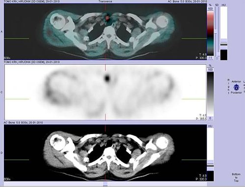
2. Scintigraphy of the parathyroid glands
Your doctor has ordered a scintigraphic examination to check for the presence of an enlarged parathyroid gland.
The examination is associated with radiation exposure, which is comparable to other X-ray examinations and which you receive in two years of your life from cosmic radiation and radiation comes from natural radioactive sources.
Preparation: Not necessary.
Design: You will receive a small amount of radioactive substance in an intravenous injection. The examination itself is performed in two steps, the first images 10 minutes after the injection, the next 1,5-2 hours. In the meantime, it will perform SPECT / CT.
1. Scintigraphic examination of the presence of Meckel's diverticulum
Principle:
Your doctor has ordered a scintigraphic examination to check that a certain area of your gut does not contain the gastric mucosa.
The examination is associated with radiation exposure, which is comparable to other X-ray examinations and which you receive in two years of your life from cosmic radiation and radiation comes from natural radioactive sources.
Preparation: Fasting 4-6 hours before the examination. Ranisan 150 mg after (in children weighing 2-4 mg / kg) in the evening and in the morning before the test. Appropriate scintigraphy interval from X-ray contrast administration for at least 48 hours.
Design: You will receive a small amount of radioactive substance in an intravenous injection. The examination itself is performed immediately after the injection and lasts 45-60 minutes, after the end you can add additional pictures lasting 3 minutes.
2. Dynamic scintigraphy of esophageal motility
Your doctor has ordered a scintigraphic examination to monitor the passage of the liquid bite through the esophagus.
The examination is associated with radiation exposure, which is comparable to other X-ray examinations and which you receive in four years of your life from cosmic radiation and radiation from natural radioactive sources.
Preparation: At least 4-6 hours of fasting, do not smoke before examination.
Design: Swallow a small amount of radioactive material. The examination itself begins with the ingestion of the substance and lasts approximately 15 minutes.
3. Scintigraphic determination of gastroesophageal reflux¬
Your doctor has ordered a scintigraphic examination to monitor for possible fluid penetration from the stomach into the esophagus.
The examination is associated with radiation exposure, which is comparable to other X-ray examinations and which you receive in four years of your life from cosmic radiation and radiation from natural radioactive sources.
Preparation: At least 4-6 hours of fasting, do not smoke before the examination. Older children and adults will bring juice or tea (0,5 liters) and the usual breakfast. Infant mothers bring one empty bottle for examination, the other with prepared milk. If breastfeeding, only one empty bottle.
Design: Swallow a small amount of radioactive material. In infants, the radioactive substance is given a small amount of milk, which the child drinks enough with milk or tea. Older children and adults receive a radiopharmaceutical in a small amount of juice or tea, they can get them on a solid diet. The examination itself begins after drinking and eating, lasting 30-60 minutes. Another image can be added in 4 hours.
4. Stomach evacuation scintigraphy
Your doctor has ordered a scintigraphic examination to monitor the progress and rate of gastric emptying after eating a semi-solid meal. The examination is associated with radiation exposure, which is comparable to other X-ray examinations and which you receive in four years of your life from cosmic radiation and radiation from natural radioactive sources.
Preparation: At least 8 hours of fasting. Diabetics bring insulin. Older children and adults will bring juice or tea (0,5 liters), a cup of milk rice (200-250 g) and two rolls or slices of bread for examination. Infant mothers bring one empty bottle for examination, the other with prepared milk. If breastfeeding, only one empty bottle.
Design: Swallow a small amount of radioactive material. In infants, the radioactive substance is given a small amount of milk, which the child drinks enough with milk or tea. Older children and adults receive radiopharmaceuticals mixed in a cup of milk rice, they can eat them with a solid diet. The examination itself starts after drinking and eating, it lasts 90 minutes. Another image can be completed in 1-4 hours.
5. Scintigraphic diagnosis of bleeding into the GIT
Your doctor has ordered a scintigraphic examination to detect bleeding into the digestive tract using marked red blood cells. The examination is associated with radiation exposure, which is comparable to other X-ray examinations and which you receive in two years of your life from cosmic radiation and radiation comes from natural radioactive sources.
Preparation: Thyroid blockade by administration of Chlorigen (performed after arrival for examination).
Design: According to the method of marking red blood cells, you will either receive a small amount of radioactive substance in an intravenous injection and the blood cells will be marked directly in your body or we will take 10 ml of blood, from which the red blood cells will separate, label with a radioactive substance. We will return the marked blood cells to you by intravenous injection. The examination itself begins with the administration of the radioactive substance, lasts 90 minutes, further images can be taken within another 24 hours.
6. Dynamic scintigraphy of the liver and bile ducts
Your doctor has ordered a scintigraphic examination to assess liver function and bile duct outflow.
The examination is associated with radiation exposure, which is comparable to other X-ray examinations and which you receive in two years of your life from cosmic radiation and radiation comes from natural radioactive sources.
Preparation: Fasting 4-6 hours before the examination. Bring dark chocolate for the test. Starvation is not necessary in infants with suspected bile duct atresia.
Design: You will receive a small amount of radioactive substance in an intravenous injection. The examination itself is performed immediately after the injection and lasts 60 minutes, after the end you can add more pictures with an interval of 30 minutes, if necessary, even longer (1-4 hours).
7. Scintigraphy of the liver and spleen
Your doctor has ordered a scintigraphic examination to show your liver and spleen.
The examination is associated with radiation exposure, which is comparable to other X-ray examinations and which you receive in two years of your life from cosmic radiation and radiation comes from natural radioactive sources.
Preparation: No need to eat, you can drink.
Design: You will receive a small amount of radioactive substance in an intravenous injection. The examination itself begins 15-30 minutes after the injection and lasts 1-2 hours.
8. Scintigraphy of the liver to detect hemangioma
Your doctor has ordered a scintigraphic examination to check for the presence of hemangiomas in the liver (a benign body made of blood vessels).
The examination is associated with radiation exposure, which is comparable to other X-ray examinations and which you will receive in two years of your life from cosmic radiation and radiation from natural radioactive sources.
Preparation: You can eat, you can drink. Thyroid blockade by administration of Chlorigen (performed after arrival for examination).
Design: We will mark your red blood cells directly in your bloodstream by giving two intravenous injections 30 minutes apart. The self-examination begins with the second injection. The first shots last approximately 30 minutes. Others are replenished in 2 hours and shooting takes 45 minutes.
Design: We will mark your red blood cells directly in your bloodstream by giving two intravenous injections 30 minutes apart. The examination itself takes approximately 30 minutes.
Palliative therapy of painful bone metastases 153Sm
What is Samarium?
Samarium is an injectable substance designed to relieve bone pain in your illness.
Samarium is chemically similar to calcium. It settles wherever the bones receive new calcium, even in painful places. Here Samarium lasts for many weeks and reduces pain. For many years, doctors have used certain types of radiation to reduce the pain to which people like you are exposed. Samarium is the result of new developments in this treatment. The injection contains a small amount of a specially selected form of radioactive Samarium, chosen so that almost all the radiation is released in the places where the Samarium binds. This allows treatment to be delivered exactly where it is needed.
Why was Samarium prescribed for me?
The substance treats bone findings and relieves pain. For many people, Samaria is much more acceptable than other treatments. It has been prescribed to you because the doctors believe that it will be the most advantageous treatment for you.
What is the procedure after injecting Samaria?
If you are an outpatient, you will be released for home care after administration. If you are hospitalized, you will return to the appropriate ward some time after application.
What effect will Samarium have?
You may temporarily increase your pain within one to three days of taking it. This is normal and the doses of sedatives can be increased in consultation with your doctor. Within about a week of application, you will experience pain relief that lasts for up to several months. Samaria may be repeated as needed.
Are there any side effects?
After serving Samaria, you can eat and drink normally. In some patients, the number of white blood cells and platelets may decrease. A decrease in white blood cells can cause infection, a decrease in platelet bleeding. Your doctor will therefore invite you for a blood test for 6 weeks after administration, the first 1-2 weeks apart.
Should I stop taking painkillers?
Your doctor may advise you to continue taking painkillers until they begin to subside. He may then recommend that you gradually reduce your dose. You can continue to reduce your doses and may not need sedatives at all. If you have any doubts, talk to your doctor.
What is the next treatment?
Your doctor will advise you on further treatment if you need it. You may have already received hormonal injections or tablets and your doctor may continue this treatment.
What activities can I run?
The injections do not stop you from doing anything you have been running. Once the effect of Samaria begins to subside by relieving your pain, you will find that you can engage in activities that were previously difficult or strenuous. There are usually no problems with this, but be careful not to overestimate your strength. If you have any further questions, ask your doctor for advice. Samarium has no influence on your ability to drive or use machines.
Who should I tell?
You should tell everyone who prescribes any treatment that you have been injected with Samaria.
What measures should I take?
The effect of Samaria within the body is limited to the painful areas in which it is concentrated. Samarium, which is not deposited in painful places, enters your blood and urine. Therefore, the following precautions should be strictly observed within 24 hours after application:
- Where a normal toilet bowl is available, use it in preference to a shell. Urinate sitting. Always rinse the toilet twice. Dry any urine-stained areas and rinse. Wear rubber gloves. Always wash your hands thoroughly after each use of the toilet.
- If your laundry is splashed with urine, wash it immediately. Work in rubber gloves. Wash the laundry separately and rinse it well.
- Store used rubber gloves in a plastic bag for 23 days in a place where your household members are not normally present, before disposing of them.
- In case of injury, rinse all blood well.
- Do not stay unnecessarily close to other people, especially children and pregnant women, for one week after administration. In case of hospitalization, the nursing staff will be instructed.
What happens when the pain returns?
If the pain returns, you should contact your doctor, who may prescribe another injection of Samaria.
Patients are hospitalized for treatment with open emitters (131I - radioiodine; 131I-mIBG) on the basis of recommendations caregiver specialists (catchment endocrinologist), sending complete documentation (operational protocol, histology, discharge report, ...) and an indicative interdisciplinary consultation at the Department of Nuclear Medicine and Endocrinology, Motol University Hospital.
The KNME inpatient department has a total of 34 beds. It is divided into a diagnostic-therapeutic part (1st bed station, located in communication node B, 1st floor), where patients with mostly lower whole-body activity are hospitalized, and a therapeutic part (2nd bed station, located in communication node B, 2st floor). , -1nd floor), where patients with higher whole-body activity are hospitalized. In the diagnostic-therapeutic part there is 8 single room, 1 double rooms, in the therapeutic station there is also 8 single room and XNUMX double rooms. The two adjoining rooms usually have shared bathroom facilities.
One single room and two double rooms at the 2nd bed station have their own bathroom and are equipped with an audiovisual monitoring system that allows staff to intensively monitor the patient and his life functions without being at the bedside. If a pediatric patient is hospitalized in the room, he or she can use the audiovisual system to communicate with the parent present in the adjoining room.
All rooms have one-way telephones, TVs and radios. There is a common room and a dining room with a TV at the diagnostic-therapeutic station. A small library located at the ward is available to patients. The way the ward works, where patients move between inpatient stations depending on the whole-body activity, does not make it possible to set up rooms with above-standard care. Paid internet access is available in the departments.
Double room on the 2nd bed station
|

The application of radioiodine tablets (in very small situations of radioiodine solution) takes place in a radioiodine laboratory equipped with a special shielded fume hood for working with open emitters. Intravenous use (131I-mIBG) take place in a single room.
Iodine application | Iodine hood |
 |  |
The operation is provided by nurses and physicians with certifications in endocrinology, internal medicine and nuclear medicine under the leadership of the head of the clinic. The Department of Radiological Physics KNME cooperates in providing diagnostic and treatment procedures using ionizing radiation sources.
Chief physician of the inpatient department of KNME:
primary MUDr. Kateřina Táborská | (224) 434 631 |
We provide information about the patient's health status to relatives (authorized) persons only with the patient's consent and only during a personal visit, not by telephone.
Diagnostic and posterior scintigraphic examinations are performed on SPECT Siemens Symbia S at the 2nd bed station. If SPECT / CT needs to be supplemented, the patient is sent to an NM clinic on the same floor. We use dose rate meters, specifically a scintillation probe, to determine whole-body activity after diagnostic application. We use dose rate meters, specifically the ionization chamber (SVLD probe), to determine whole-body activity after therapeutic application.
| Symbia S | Symbia S |
 |  |
The endocrinology outpatient clinic is located in communication node A, 3rd floor. It provides consulting services for individual departments of the Motol University Hospital in the entire scope of the field, but patients are also treated in a network of outpatient outpatient endocrinologists. The main task of the department is to ensure the treatment of patients from the Czech Republic with differentiated thyroid carcinoma and moderate and severe thyroid orbitopathy. The department works closely with the Department of Internal Medicine and the Department of Clinical Biochemistry in the treatment of patients with metabolic disorders and with the Department of Gynecology and Obstetrics, Charles University, 2nd Faculty of Medicine in the treatment of sterility. Provides sonographic thyroid diagnostics, including targeted thin needle aspiration biopsy (FNAB). After a conciliar examination (except in the most serious cases), the patients are transferred to the catchment endocrinological facilities.
Examinations are performed on the recommendation of a specialist from the Motol University Hospital or a certified endocrinologist by prior telephone order.
Head of KNME:
| prof. MUDr. Petr Vlcek, CSc., MHA | (224) 434 600 | petr.vlcek@fnmotol.cz |
Chief physician of the KNME endocrinology outpatient clinic:
| MD Kateřina Personová | (224) 434 628 |
Doctors:
| MD Ondřej Hádek | (224) 434 628 | ondrej.hadek@fnmotol.cz |
| MD Lucie Lančová | (224) 434 628 | lucie.lancova@fnmotol.cz |
| MUDr. Magdalena Matejkova Behanova, PhD. | (224) 434 628 | magdalena.matejkova@fnmotol.cz |
| MD Olga Nývltová | (224) 434 628 | olga.nyvltova@fnmotol.cz |
| MD Pavel Racek | (224) 434 628 | pavel.racek@fnmotol.cz |
| MD Veronika Simonová | (224) 434 628 | veronika.simonova@fnmotol.cz |
| MD Pavla Sýkorová | (224) 434 628 | pavla.sykorova@fnmotol.cz |
| MUDr. Květuše Vošmiková, Ph.D. | (224) 434 628 | kvetuse.vosmikova@fnmotol.cz |
Office hours
Ordering examinations on the phone number: (224) 434 628
We change your appointment changes or other requirements by phone - for operational reasons - only on working days between 7.00 - 10.00. Thank you for your understanding.
Reorder dates and e-recipes: lenka.andelova@fnmotol.cz, martina.verner@fnmotol.cz
Warning:
We provide endocrinological conciliation examinations only within the Motol University Hospital or, upon agreement of the superconsiliary service, at the written request of a certified endocrinologist within the Czech Republic. We dispensary patients with differentiated and medullary thyroid carcinoma and severe forms of thyroid orbitopathy, we are not able to treat other endocrine diseases for capacity reasons and we refer these patients to standard field endocrinology departments for dispensary care.
Surgery hours only for booked patients (valid from January 2018):
| Hádek O., MD | Tuesday 8:00 - 12:30 |
| Hollay E., M.D. | Tuesday 8:00 - 10:00 |
| Křenek M., M.D., CSc. | Tuesday 10:00 - 12:00 |
| Lančová L., MD | Friday 8:00 - 12:30 |
| Matějková Běhanová M., MD, PhD. | Monday 8:00 - 14:00 |
| Nývltová O., MD | Thursday 9:00 - 13:00 |
| Personova K., MUDr. | Monday 8:00 - 12:00 |
| Racek P., MD | Monday 8:00 - 12:00 |
| Simonová V., M.D. | Wednesday 8:00 - 13:00 |
| Sykorova P., MUDr. | Thursday 9:00 - 12:00 |
| Vlček P., prof., MD, CSc., MHA | Tuesday 8:00 - 14:00 |
| Vosmikova K., MUDr., PhD. | Wednesday 8:00 - 14:00 |
Blood collection:
Monday to Wednesday 7:00 - 14:00
Thursday 8:45 - 14:00
Friday 7:00 - 14:00
The endocrinology outpatient clinic is equipped with Toshiba Nemio SSA-550A / E and Toshiba SSA-250A sonographers
Toshiba Nemio SSA-550A / E
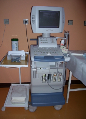
Toshiba SSA-250A
Radiological physics and radiation protection
The issues of radiological physics and radiation protection are handled for the KNME by the employees of the Independent Department of Medical Physics - nuclear medicine section. This is a separate department, which is located in the premises of the clinic in the section "KNME - Clinic management" in communication node B, -2. floor. Clinical radiological physicists, radiological physicists and radiological technicians work in the department.
Department:
- ensures regular quality control of imaging systems and measuring technology
- ensures the application and optimization of radiation protection in the provision of health care, especially radiation protection of patients during medical exposure, radiation protection of workers, workplaces and their surroundings
- ensures continuous monitoring of compliance with radiation protection requirements
- determines the radiation exposure of the patient from examination or treatment with a radionuclide
- cooperates with physicians in the acquisition and evaluation of clinical data
- cooperates in the introduction of new diagnostic and therapeutic methods in accordance with the recommendations of the European Association of Nuclear Medicine (EANM)
- ensures compliance of documentation and records of the workplace with legislative regulations and recommendations of the State Office for Nuclear Safety concerning the management of ionizing radiation sources and radiation protection
- participates in the solution of scientific grants
- participates in teaching within KNME
Clinical radiological physicist
Ing. Dana Prchalová (head of SOLF-NM) | (224) 434 611 | |
Ing. Tereza Kračmerová | (224) 434 612 |
Radiological Physicist:
Ing. Alena Kenová | (224) 434 612 |
Radiology technician:
Bc. Tomáš Řeháček | 224 | |
Bc. Sonia Burešová | 224 |
- Projects
- Publications
- Grant agencies
- Teaching
Doc. MUDr. Karel Šilink, DrSc., The disciple of the founder of Czech endocrinology, academic prof. Josef Charvát, DrSc. The workplace was established as a detached bed base of the Research Institute of Endocrinology (VÚE) founded by Šilinka and was ceremoniously opened on 1 July 7.
doc. MUDr. Karel Silink, DrSc.

Until then, it was not possible to administer therapeutic doses systematically 131 Also to patients with thyrotoxicosis and thyroid cancer (the first experimental therapies in our country were given in 1956-9 at the VÚE and hospitalization took place internally at the hospital in the Old School in Dušní Street). The capacity of 20 beds was used for diagnostics and, since January 1960, also for therapy with open emitters, the focus of the work of the inpatient department was mainly in the therapy of thyrotoxicosis. After equipping the workplace with a Siemens scintigrapher (1962), it was possible to expand the services with a consultation examination for the Municipal Hospital in Prague 5 Motol and the workplace was partially integrated into the structures of the MěN in Motol as a cooperating internal department. They consisted of about 30 employees from VÚE and Motol Hospital, the department was headed by MUDr. Jan Kubal. Around 400 patients a year were examined in the outpatient clinics and beds. Before 1973, before the era of cytostatics, about 200 patients were treated by intracavitary application of radiogold colloid in cooperation with the gynecological department. 198Au. Close cooperation with the head of the surgical department prof. MUDr. B. Niederlem, DrSc. an excellent thyroid surgeon, enabled the introduction of so-called "hot" operations on thyroid tumors after the application of radioiodine.
From 1965, the Radioisotope Department was headed by MUDr. Jan Nemec, CSc. and the main area of interest has shifted to the treatment of differentiated follicular and papillary thyroid carcinoma, this line of work continues to this day.
prof. MUDr. Jan Nemec, DrSc.

Further development of diagnostics and therapy took place in 1972-1974 with the installation of the Picker color scintigrapher and the extension of the second pavilion, where the diagnostic part of the radioisotope department was moved, which increased the bed stock capacity to 25 and after further reconstruction in 1980 to 29 beds.
In 1970, a specialized clinic for the treatment of severe forms of endocrine orbitopathy was established, headed by MUDr. Stanislav Váňa, CSc., Who laid the foundations for the emergence of interdisciplinary cooperation between an endocrinologist and an ophthalmologist in one surgery.
In addition to the treatment and nationwide dispensary of patients with papillary and follicular thyroid cancer (there is a therapeutic gradient for most of Slovakia at that time), the workplace has been working since 1978, under the leadership of MUDr. Marta Neradilová, CSc., Also began to focus on nationwide care for patients with medullary thyroid cancer.
In 1985, the Department of Nuclear Medicine in Motol was established in connection with the change of the hospital's statute to the University Hospital in Motol. This department, together with the inpatient department of the VÚE, became the basis for the newly established Department of Nuclear Medicine (1992), a joint workplace of the 2nd Faculty of Medicine, Charles University and the University Hospital in Motol. MUDr. Jan Nemec, DrSc.
Major changes at the clinic occurred by moving to the new premises of the blue pavilion of the University Hospital in Motol in March 1998. The clinic acquired modern equipment and premises worthy of workplaces of national importance. This enabled the further development of nuclear diagnostics in particular, with a number of special radionuclide examinations and new treatments for non-thyroid diseases. From the same year, the KNM radioimmunoassay laboratory became a part of the Institute of Clinical Biochemistry, Charles University, 2nd Medical Faculty and Motol University Hospital.
In 1999, he replaced prof. German doc. MUDr. Petr Vlcek, CSc. Under his leadership, basic diagnostic and treatment programs are further developed, cooperation with other clinics is expanding, and rich scientific research and pedagogical activities continue.
The clinic currently has around 60 employees. The clinic's registry consists of 45 patients; at the end of 000, more than 2010 patients with differentiated thyroid cancer were treated (this is one of the largest groups in the world). Around 11300 patients (including 950% of cancers) are hospitalized in the inpatient department every year. Around 95 patients are examined in the clinic's endocrinology clinic and around 13 patients a year in the nuclear medicine clinic.
The research activities of the clinic are closely connected with clinical practice. In the 90s, scintigraphic diagnostics was introduced into the standard examination program 99mTc-MIBI. The first therapies took place at the clinic 131I-mIBG in patients with medullary thyroid carcinoma (1994), in collaboration with the Department of Pediatric Oncology, Charles University, 2nd Faculty of Medicine in children with malignant neuroblastoma (1997), carcinoid (1999) and malignant pheochromocytoma (2001).
In 2001, by the director of the University Hospital in Motol, the Center for Thyroid Oncology and Thyroid Orbitopathy of the Charles University 2nd Medical Faculty and the University Hospital Motol was established at the clinic, under the leadership of doc. Vlčka, which provides professional counseling care for the entire Czech Republic. In 2002, by decision of the Minister of Health of the Czech Republic, the workplace was renamed, in accordance with the scope of medical-preventive activities and the focus of undergraduate and postgraduate teaching, at the Department of Nuclear Medicine and Endocrinology, Charles University, 2nd Medical Faculty and Motol University Hospital (KNME).
Since 2008, KNME has been an approved center for the treatment of oncological diseases using a radiopharmaceutical 131I-mIBG, diagnostics and therapy after preparation with human recombinant thyroid stimulating hormone (rhTSH) and for the treatment of severe thyroid autoimmune orbitopathy (TAO) somatostatin analogues.
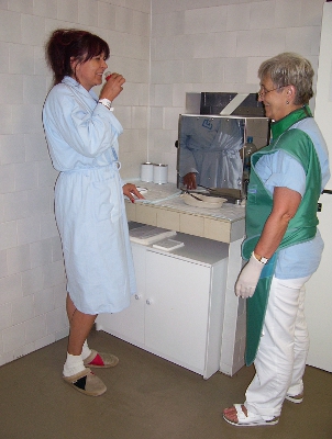
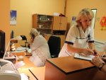 Reception of patients - ambulance

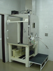
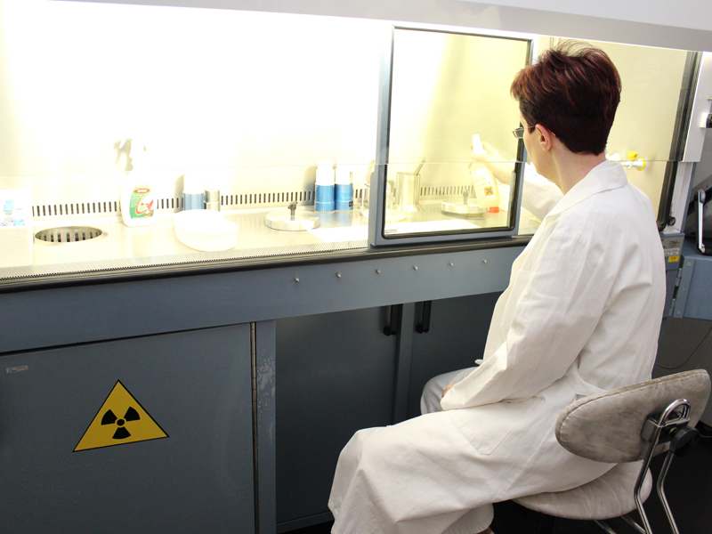 Production of radiopharmaceuticals
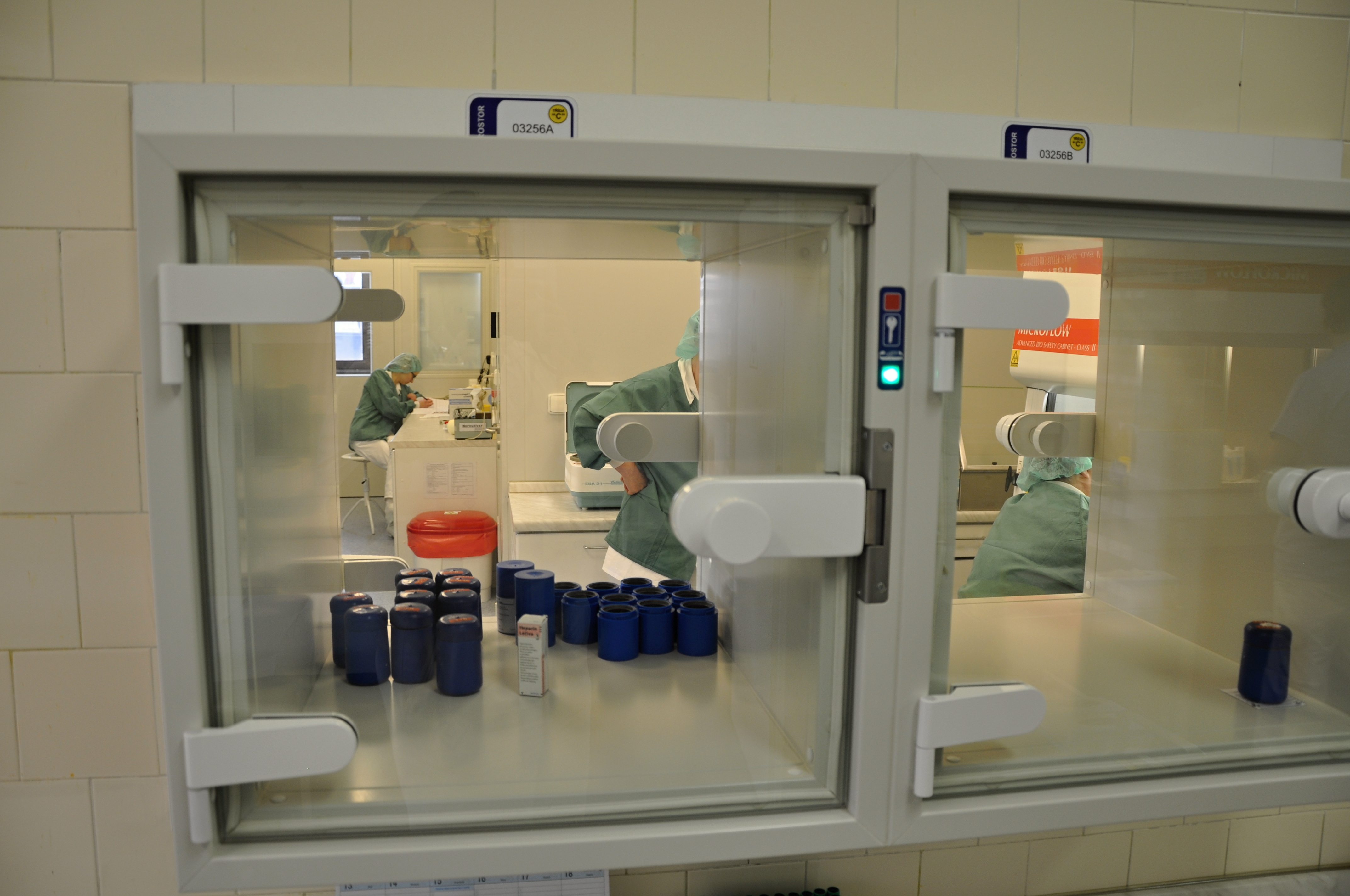 Release from pharmacological laboratory
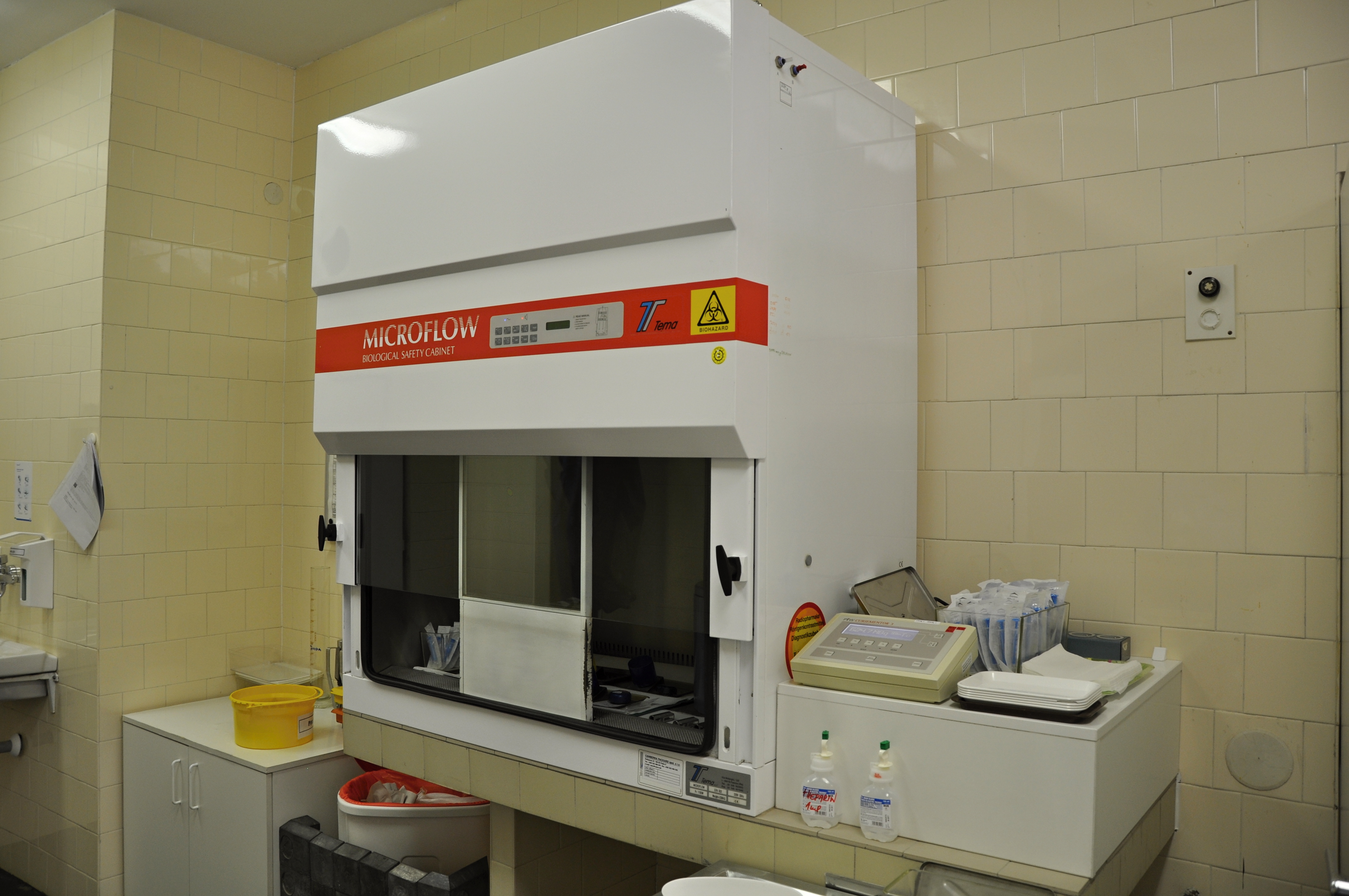 Application box
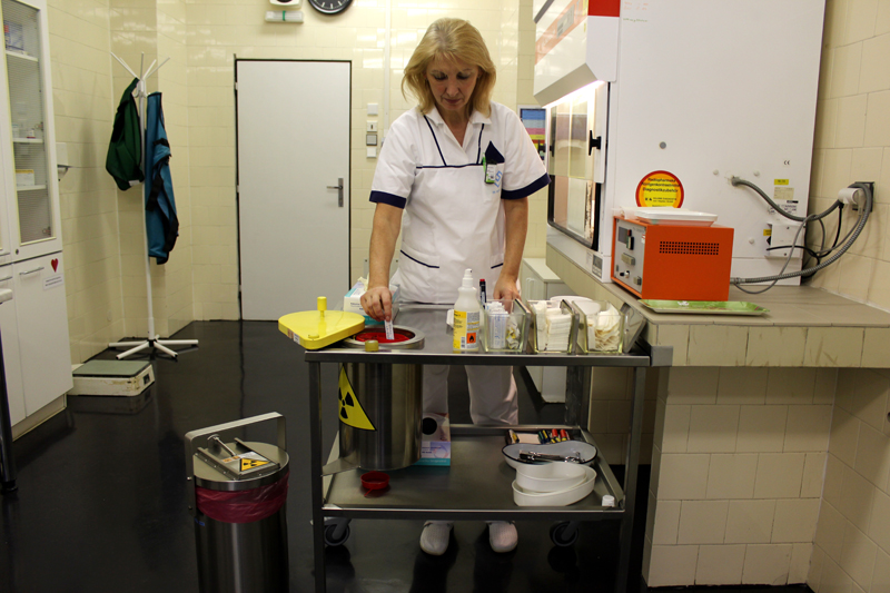 Preparation of a radiopharmaceutical for a given patient
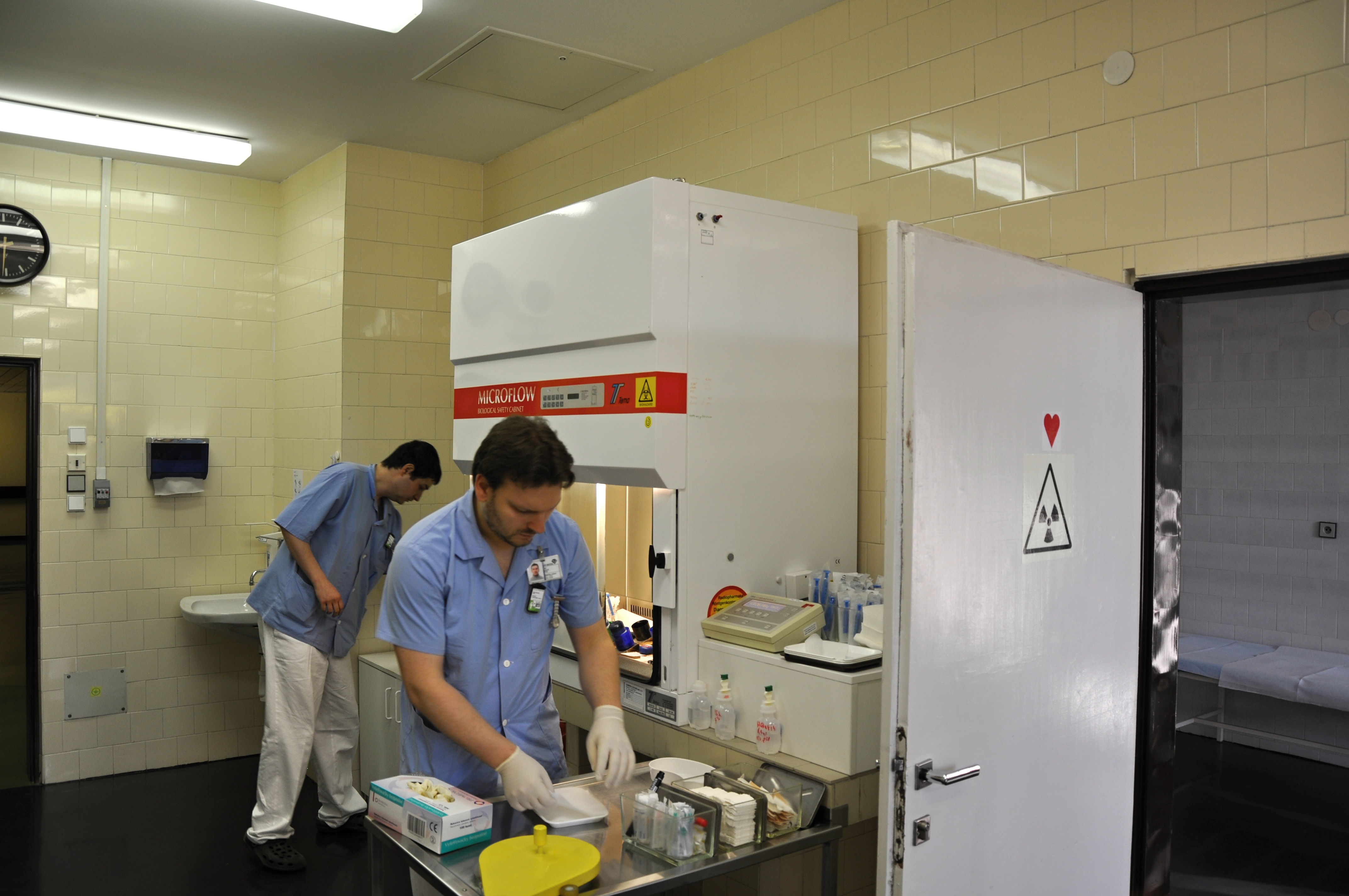 Preparation of a radiopharmaceutical for a given patient
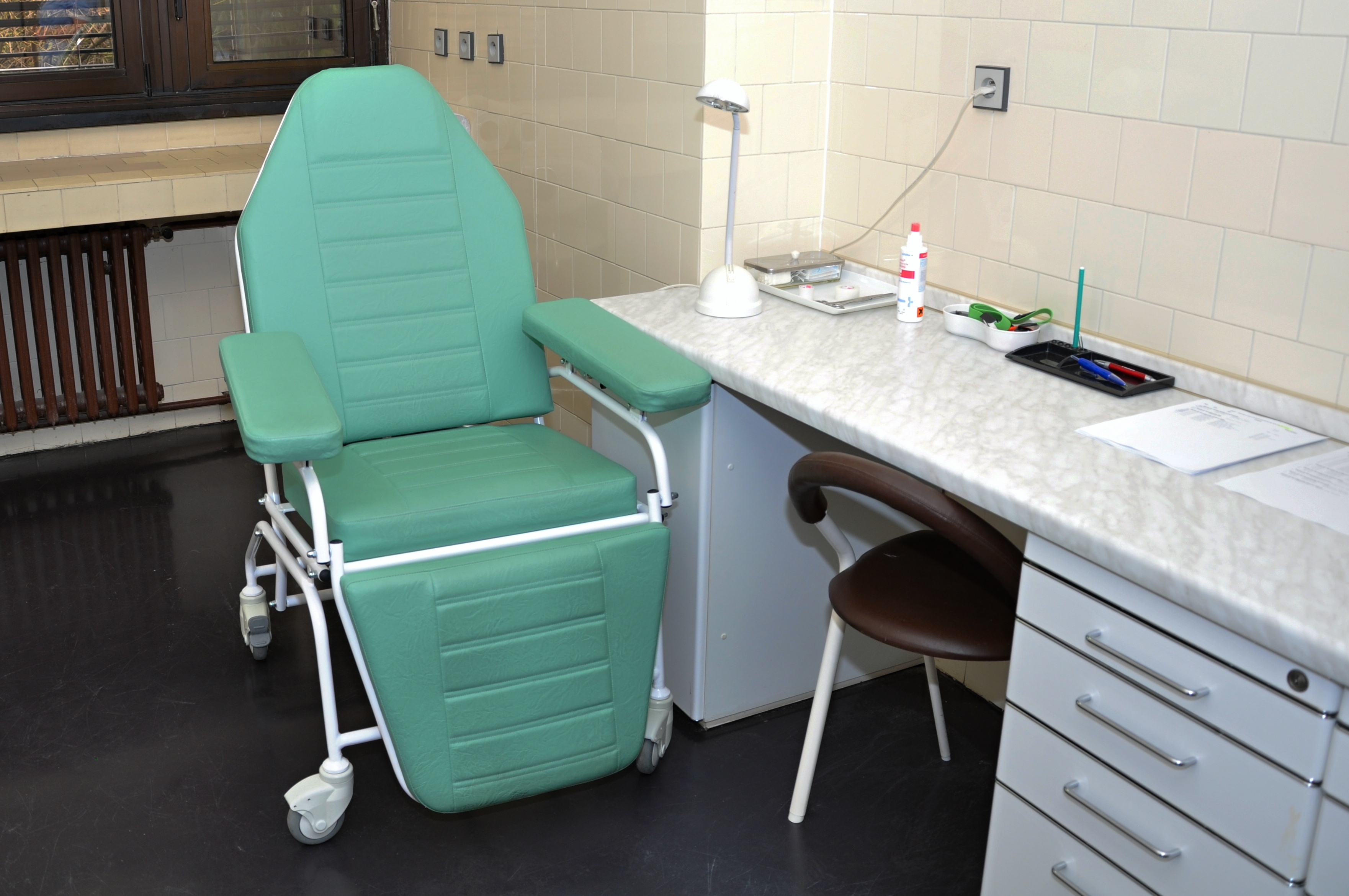 Application chair
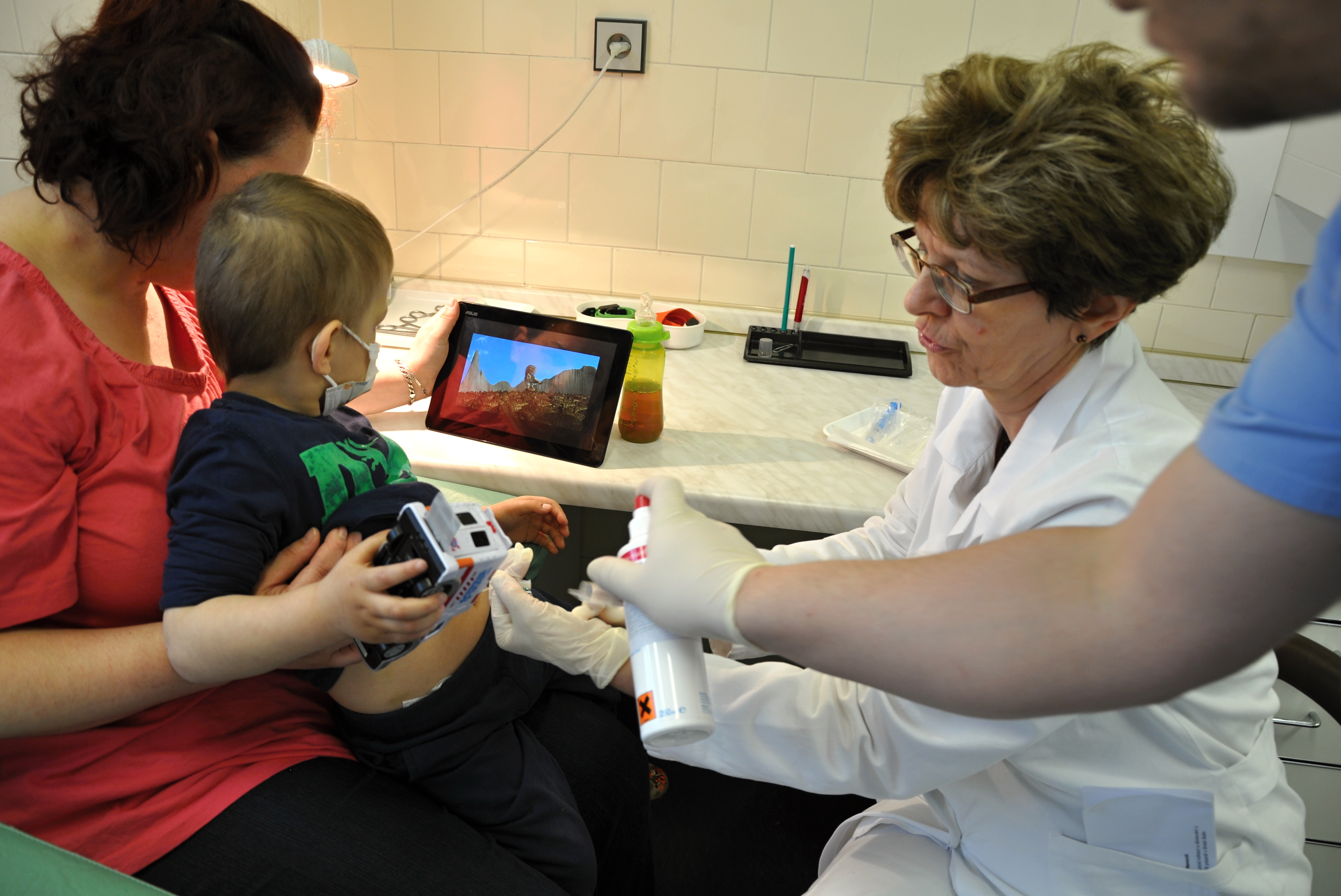 Application
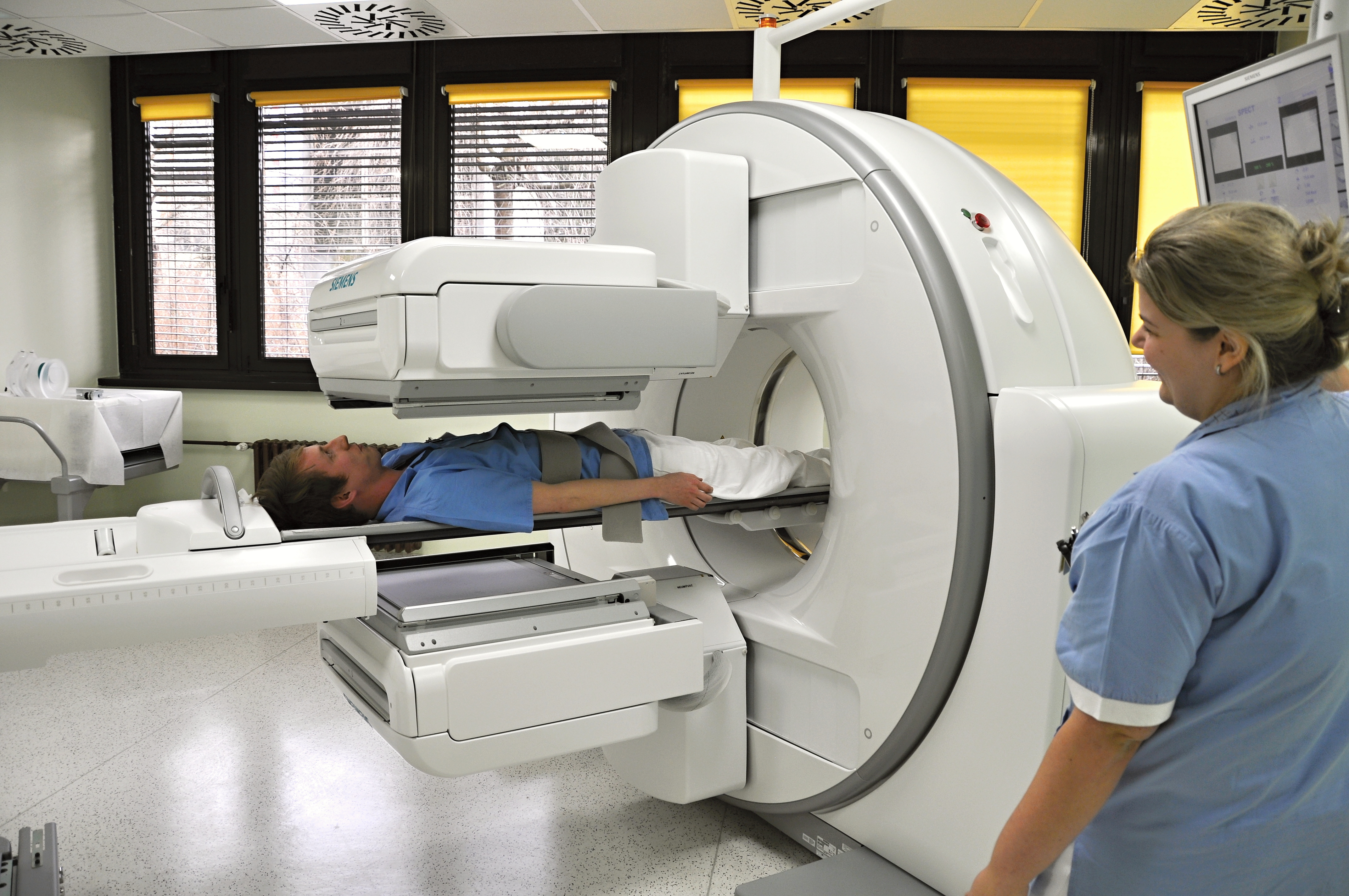 Scintillation camera SPECT / CT Siemens Intevo ...
 Scintillation camera SPECT / CT Siemens Intevo ...
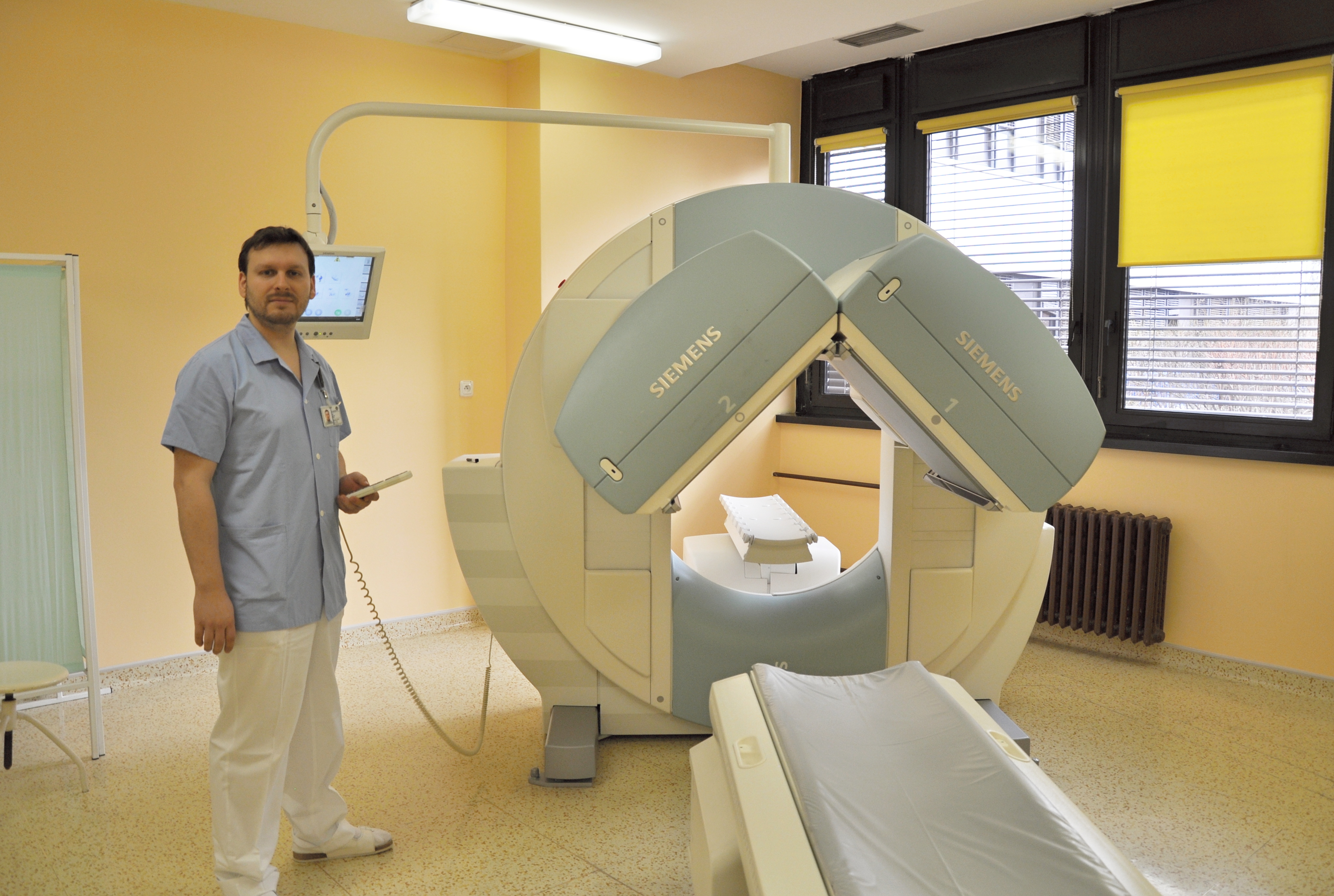 SPECT Siemens Symbia S scintillation camera
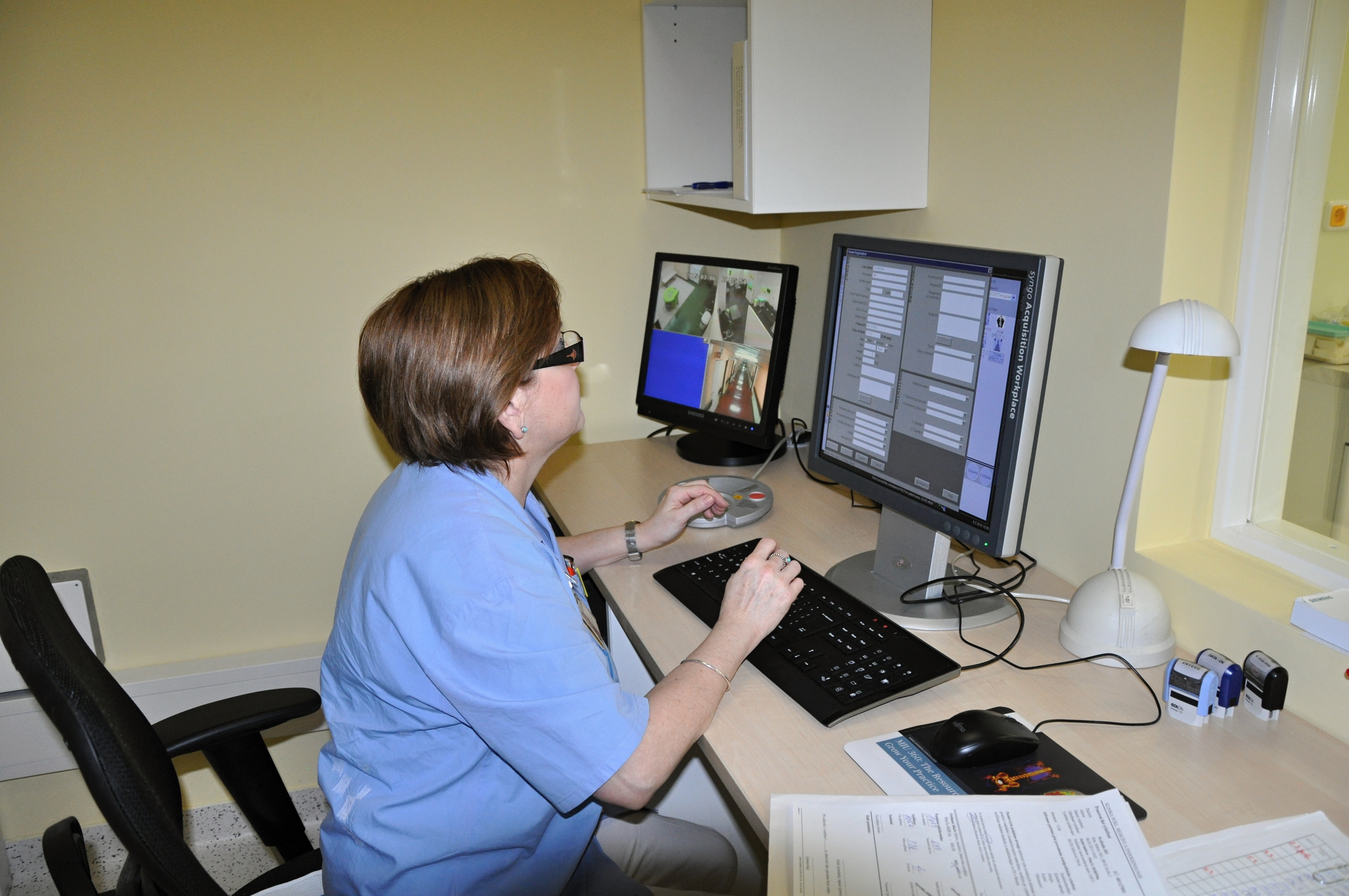 SPECT / CT control room
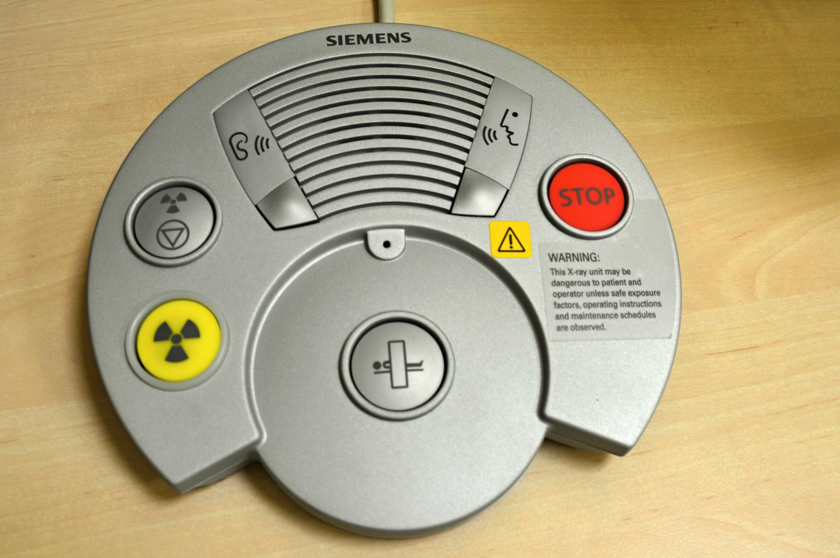 SPECT / CT control room
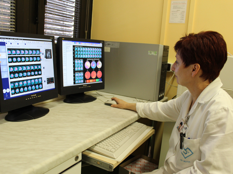 Image evaluation
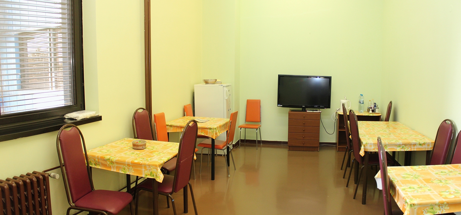 Inpatient department
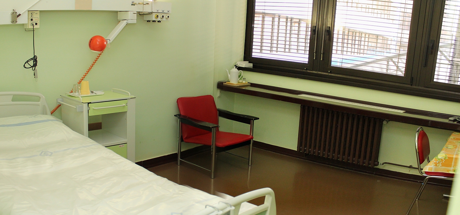 Inpatient department
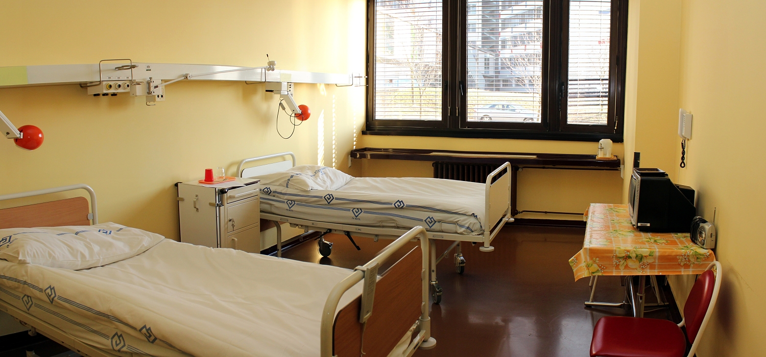 Inpatient department
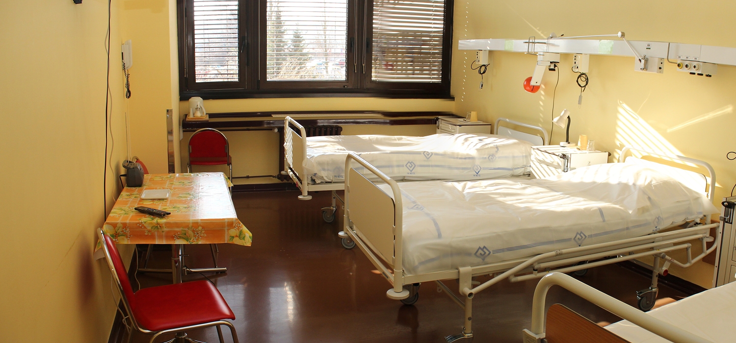 Inpatient department
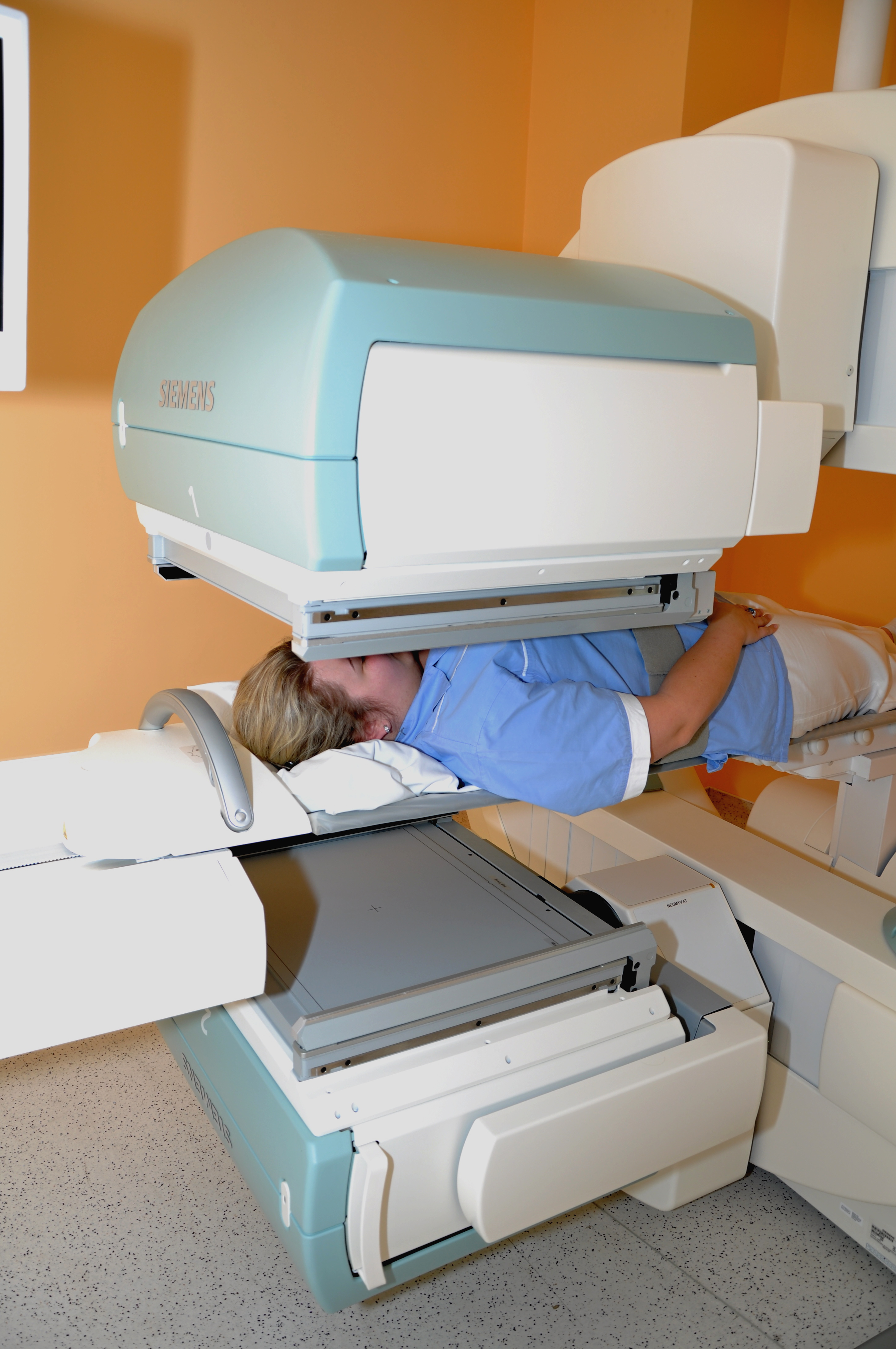 Scintillation camera SPECT Siemens Symbia S -...
 Scintillation camera SPECT Siemens Symbia S -...
Contact
The head
prof. MUDr. Petr Vlcek, CSc., MHA
personal website
phone: 224 434 600
Email: petr.vlcek@fnmotol.cz
Secretariat of the Head
Petra Luptáková
phone: 224 434 601
Email: nuclear@fnmotol.cz
Lenka Houserová
phone: 224 434 602
fax: +420 224 434 620
Email: nuclear@fnmotol.cz
Deputy Head for Medical-Preventive Care - Head of the Clinic
MD Kateřina Táborská
phone: 224 434 626
Email: katerina.taborska@fnmotol.cz
Deputy head for pedagogical activities
MD Lucie Lančová
phone: 224 434 667
Email: lucie.lancova@fnmotol.cz
Head nurse
Ladislava Novotná
phone: 224 434 605
Email: ladislava.novotna@fnmotol.cz
Endocrinology clinic
Endocrinology clinic reception
phone: 224 434 628
Chief physician of the endocrinology clinic
MD Kateřina Personová
phone: 224 434 628
Email: katerina.personova@fnmotol.cz
Nuclear medicine clinic
Nuclear medicine clinic reception
phone: 224 434 626
Chief physician of the nuclear medicine clinic
MD Kateřina Táborská
phone: 224 434 626
Email: katerina.taborska@fnmotol.cz
Inpatient department
Chief physician of the inpatient department
MD Kateřina Táborská
phone: 224 434 626
Email: katerina.taborska@fnmotol.cz
Inpatient department
Nurse 1st station, tel .: 224 434 631
Nurse 2st station, tel .: 224 434 630
Laboratory of therapeutic radiopharmaceuticals, tel .: 224 434 627
Radiopharmaceutical laboratory
Head of the radiopharmaceutical laboratory
RNDr. Martin Vlk, Ph.D.
phone: 224 434 665
Email: martin.vlk@fnmotol.cz
Separate department of medical physics – nuclear medicine section
Head of SOLF-NM department
Ing. Dana Prchalová
phone: 224 434 611
Email: dana.prchalova@fnmotol.cz
Supervisor
MD Kateřina Táborská
phone: 224 434 626
Email: katerina.taborska@fnmotol.cz