Due to capacity reasons, it will not be possible to take pictures of patients with a request issued elsewhere than at Motol Hospital.
Due to capacity reasons, it will not be possible in the children's section of the KZM to examine patients with a request issued elsewhere than in Motol FN.
In addition to special examinations:
- bone age
- scanography of the lower limbs
- long format spine images
Basic information
Email requests for sending images to ePACS to: epacs@fnmotol.cz
Operating hours for ambulatory operation: Mon-Fri 7:00 - 15:00
The adult part of KZM performed a total of 2021 examinations in 306, of which:
- skiagraphic and skiascopic examinations – over 154
- contrast fluoroscopic examination of the alimentary canal and urogenital tract – 1120
- Ultrasound examination – around 9
- CT scan – over 25
- MR examination – around 10
In 2021, the children's section of the KZM performed a total of 57 examinations, of which:
- skiagraphic and skiascopic examination – 35783
- Ultrasound examination – 15
- CT scan – 2504
- MR examination – 3417
- dental CT – 495
History of the clinic
The X-ray department at the FN in Motola was founded and led for a long time by one of the pioneers of Czech radiology, Prof. MD Slavoj Věšín, DrSc. After the opening of the monoblock of the children's section of the Motol FN in 1978, she headed the radiodiagnostic department doc. MD Eva. Kolihová, CSc. Under her leadership, the department became a leading center for pediatric radiology in our country. In 1990, prof. became the head of the radiodiagnostic department. MD Stanislav Tůma, CSc., who in 992 succeeded in obtaining the status of a clinic for the department. In 997, the construction of the blue pavilion was completed, and the Imaging Methods Clinic became a top-level workplace thanks to its equipment with the most modern, powerful devices. In the years 1999 - 2007, the head of the Imaging Methods Clinic was prof. MD Jiří Neuwirth, CSc. Since October 2007, the head of the Imaging Methods Clinic is prof. MD Miloslav Roček, CSc. The clinic of imaging methods has become a fully digitized workplace, most of the devices have been replaced with new top-of-the-line devices. A transcription of the dictated word for each doctor was put into operation. The workplace currently enables the comprehensive implementation of all diagnostic and therapeutic procedures for children and adult patients. In autumn 2011, a new children's diagnostic center was opened, which is fully equipped, including a CT and MR machine. This workplace is currently one of the most modern workplaces of its kind in Europe.
Ordering patients (records):
Tel.: 224 438 133, 224 438 124
Fax.: 224 438 125
THE ADULT PART
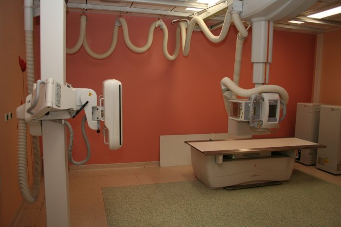
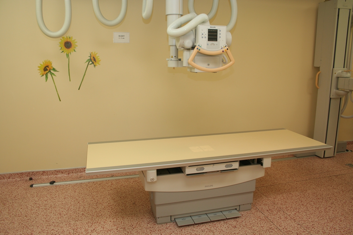
- Skiascopic-skiagraphic department for adults
- Bone diagnosis of adults
- Adult Computed Tomography (CT)
- Adult Magnetic Resonance Imaging (MR)
- Sonography for adults (UZ, USG)
- Breast mammography and sonography
- Interventional radiology
- Whole body densitometry
CHILDREN'S PART
- Pediatric conventional radiology
- Pediatric Computed Tomography (CT)
- Pediatric sonography (UZ, USG)
- Department of Pediatric Magnetic Resonance (MR)
Skiaskopicko-skiagrafické odd. for adults
Ordering patients for fluoroscopy examinations:
Phone: 224 438 183
Chief doctor
MD Michal Polovinčak
Tel.: 224 438 153
Email: michal.polovincak@fnmotol.cz
For the year 2021, the department performed about 31 fluoroscopy examinations and 000 fluoroscopy examinations of the alimentary canal, urogenital tract and interventional procedures. The fluoroscopy workplace is equipped with two folding walls. The skiagraphic workplace is fully digitized.
Skiagraphic and skiascopic examinations work with X-ray radiation that penetrates the tissues of the human body and enables the visualization of deeper anatomical structures. An examination with a contrast material is used to make visible structures that are not visible on a classic image - hollow organs, blood vessels.
Preparation before examination:
X-ray of the act of swallowing, passage through the upper GIT - Do not eat, drink or smoke 8 hours before the examination
X-ray irrigography of the colon
- Preparation with Fortrans according to the instructions of the referring physician.
- Do not eat, drink or smoke from midnight before the examination.
- If you have been given Buscopan during the examination, you may have temporary blurred vision or dizziness, in which case you cannot drive until these symptoms subside. It is more convenient to arrive for the examination by public transport or with a private transport.
X-ray defecography
- Preparation with Fortrans according to the instructions of the referring physician.
- Do not eat, drink or smoke from midnight before the examination.
- Before the examination, you will be given a barium contrast agent to drink for better visualization of the loops of the small intestine during the examination. Therefore, arrive well in advance (at least 30 minutes).
Urethrocystography (UCG) – no preparation is necessary, ideally an empty bladder
Diaphragm fluoroscopy – no preparation is necessary
Whole body densitometry
Ordering patients (records):
Tel.: 224 438 133, 224 438 124
Instrumentation:
Since 2019, our department has been equipped with the latest Hologic HORIZON A device for measuring BMD in adults and children, both inpatients and outpatients.

It is a whole-body densitometer equipped with a rotating arm. Among the significant innovations compared to the previous series are detector arrays with gadolinium-sulfoxylate sensors, which are characterized by a high signal-to-noise ratio and thus ensure high image quality, especially for applications that use only one X-ray beam energy for scanning. These devices meet all requirements for both clinical examinations and scientific research and respect the latest trends in bone densitometry. They are characterized by high accuracy and repeatability of measurements (less than 1% in the sense of "precision" and "accuracy"), low radiation load and high comfort for the patient and the operating staff.
The device can measure BMD on the L spine, femurs, forearms and the whole body. In children, it is possible to recalculate BMD to bone age. In addition to osteoporosis, we measure the composition of body tissues, BMI, FMI, VAT (prediction of the amount of visceral fat). In addition to these basic measurements, it is possible to take a lateral image of the thoracic spine in the supine position without positioning the patient, take atypical femur fractures and measure BMD around the TEP. Added value when measuring osteoporosis is the equipment of the device with software for measuring TBS and HSA, which refine the search for patients with an increased risk of osteoporotic fractures.
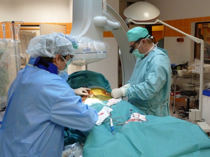
Interventional radiology
Ordering patients (records):
Tel.: 224 438 178
Chief doctor
MD Radek Pádr, MHA
Tel.: 224 438 157, 224 438 100
Email: radek.padr@fnmotol.cz
Performances performed:
- The entire spectrum of endovascular procedures on the entire arterial and venous system, including the portal tract, except for the heart. Both adult and pediatric patients are treated.
- The most common endovascular procedure is percutaneous transluminal angioplasty (PTA) of the lower limbs and visceral arteries. Furthermore, mechanical and aspiration thrombectomy for pulmonary embolism or embolic CMP, local thrombolysis of arteries and veins, treatment of vascular hemodialysis approaches, embolization for bleeding (GIT, hemoptysis, post-traumatic and iatrogenic injuries).
- As the only workplace, we routinely perform endovascular solutions for chronic venous occlusions.
- Combined procedures in cooperation with the Department of Cardiovascular Surgery of the 2nd Faculty of Medicine of the UK and the Motol Medical Center.
- Non-vascular interventions throughout.
Doctors of the angiography department give lectures at national and international congresses, which they also organize and co-organize.
Computed tomography for adults
Email requests for sending images to ePACS to: epacs@fnmotol.cz
Ordering patients (CT records):
Tel.: 224 438 185
Chief doctor
MD Radim Pavlik
Tel.: 224 438 186
E-mail: radim.pavlík@fnmotol.cz
Examinations performed:
In 2021, the department performed over 25 examinations.
The entire spectrum of diagnostic and interventional/therapeutic procedures for hospitalized and outpatient patients. In addition to common examinations such as CT of the head, neck, chest, abdomen and small pelvis, spine and limbs, we also perform specialized examinations such as CT of the heart, CT coronary angiography, CT colography, CT virtual bronchoscopy, dental CT.
The department works closely with the pulmonology and surgery clinics to monitor patients before and after lung transplantation.
Among the interventional procedures, we perform root injections of the lumbar and cervical spine (PRT), biopsy sampling for histological examination, drainage of fluid collections and pneumothoraxes, tumor ablation (e.g. RFA), chemical sympathectomy.
Preparation before examination:
- Do not drink, eat or smoke 4 hours before the CT examination with the administration of contrast material.
- Drink plenty of fluids the day before and after the examination.
- Before CT colonography, it is necessary to administer a preparation to empty the large intestine (see CT colonography).
Machines:
- Siemens Somatom Force
- Toshiba Aquilion One Next Generation
- Toshiba Aquilion Prime
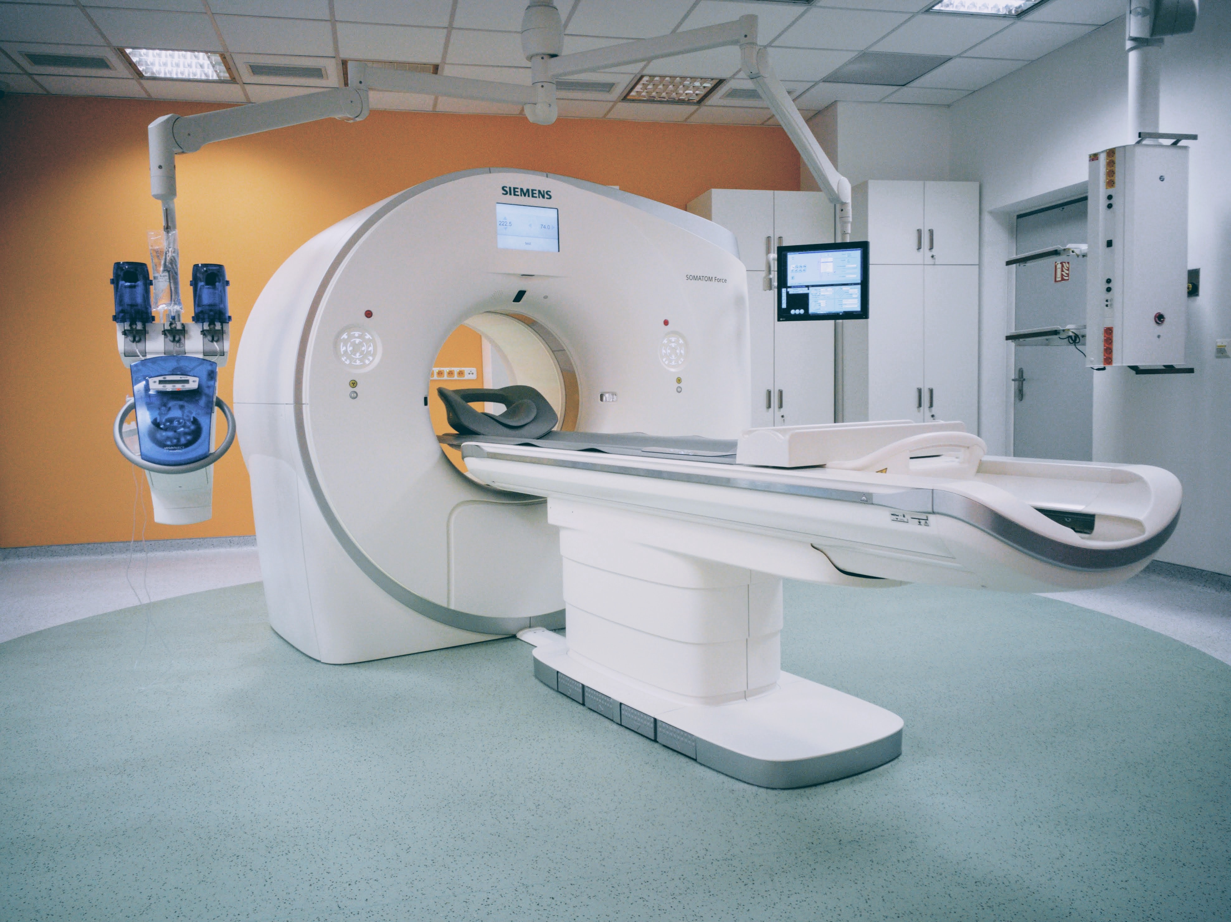
Photo gallery CT adult
 CT Aquilion Prime
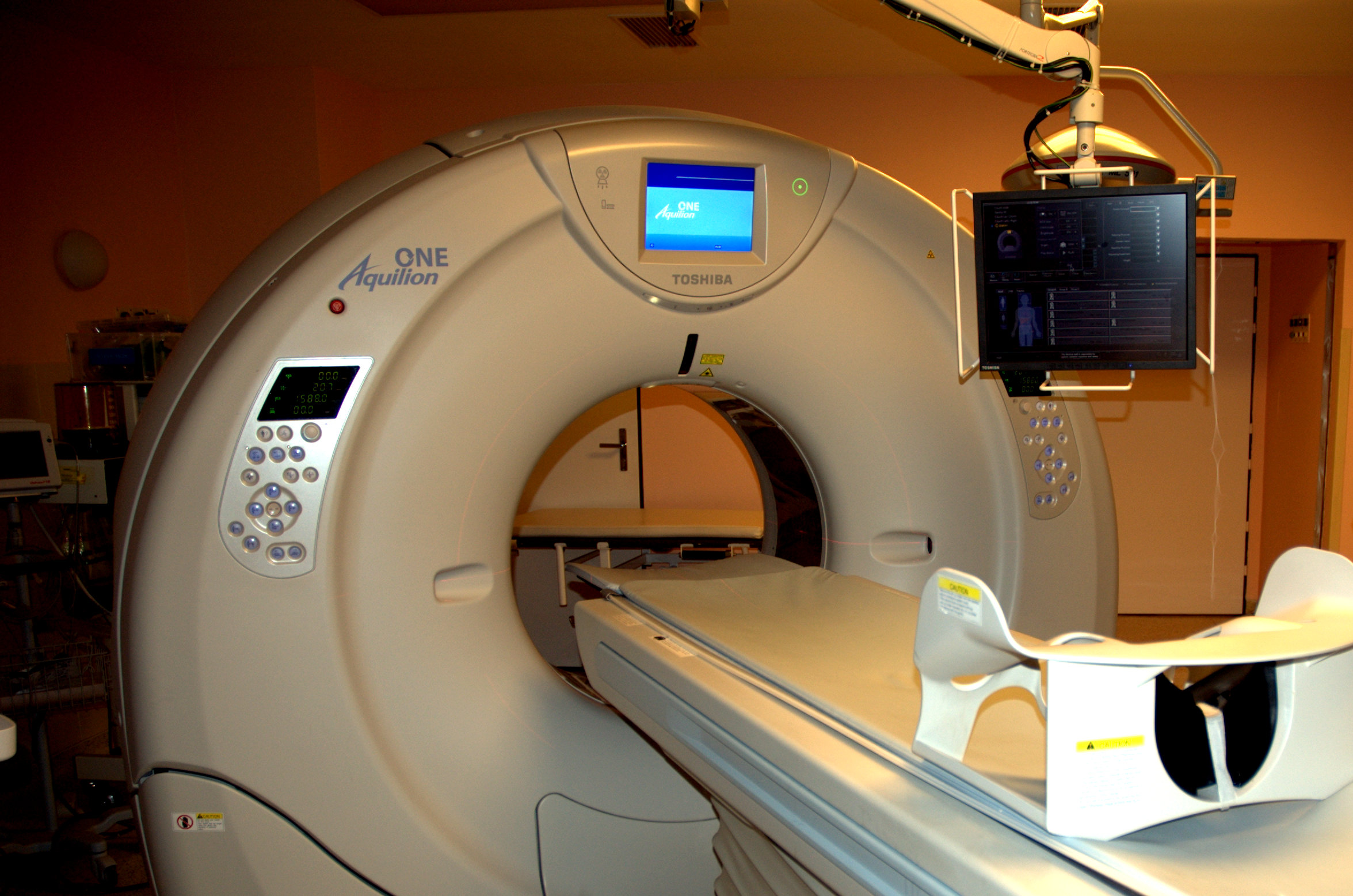 CT Aquilion ONE
 CT Somatom Force
CT coronarography
Ordering patients (CT records):
Tel.: 224 438 185
CT coronary angiography is an examination that enables the visualization of the state of the heart's arteries using computed tomography (this is X-ray radiation) with the use of an iodine contrast agent.
The heart is a constantly moving organ, and to achieve optimal examination quality we need to eliminate unwanted irregularities in its movement. To visualize the heart's arteries, it is necessary to reduce the heart rate below 60 beats per minute. In some cases, after considering all the circumstances, it may therefore be necessary for you to inject a medicine to reduce the heart rate (slow down the heart) from the group of Beta-blockers. To increase the lumen of the heart arteries, you will be administered the medicine Nitroglycerin in a spray - 2 sprays under the tongue.
Preparation before examination:
Preparation the day before the examination:
- Avoid consuming drinks containing caffeine (coffee, tea, cola) and eating chocolate
- Take care of your drinking regime (unsweetened, non-carbonated drinks)
Preparation on the day of the examination:
- Do not eat for 4 hours before the examination, drinking only clean still water is allowed
- Avoid intense sports activity on the day of the examination
Preparation for women:
- If you suspect that you might be pregnant, do not undergo the examination and cancel the appointment
- If pregnancy is not confirmed, reschedule for another date (the best date is within the first 10 days from the expected start of menstruation).
Preparation for men:
- If you are taking drugs for erectile dysfunction (e.g. Viagra and similar) - do not take these drugs 24 hours before and the following two days after the examination due to the possible unwanted cumulation of the drug's effects on the heart
Taking other medicines for women and men:
- If you are taking Metformin, ask your doctor for a kidney function test and come to the CT scan with its results.
- If you suffer from an allergy to iodinated contrast material, thyrotoxicosis, severe aortic stenosis, heart failure or AV block, inform the staff at the CT office immediately.
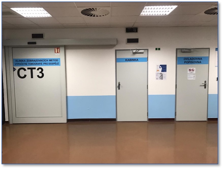
Where to find us:
- Arrive at the department of the Imaging Methods Clinic (blue building, node A, floor minus 1) 30 minutes before the examination.
- With a request form from your doctor, come to the KZM register, where you will be registered in the information system. Then wait in the waiting room across the main corridor - it is marked CT3.
Before the examination itself, you will receive an informed consent for the examination and a questionnaire from our staff to review, which please read and fill out carefully. The doctor performing the examination will answer any questions you may have.
Examination procedure:
- The nurse will invite you to the cabin to prepare for the examination. When prompted, you will undress halfway in the cabin and remove the metal ornaments from your neck and chest.
- You will lie on your back on the examination table. We will insert a cannula into a vein on your upper limb and connect the ECG leads to your chest. Intravenous access is necessary for the application of a contrast agent and also in case of the need to administer a drug to slow down the heart rate. During the entire examination, we will monitor your heart activity.
- We will monitor you through the glass from the examination room throughout the examination. The device has a built-in two-way communication device that allows you to talk to us at any time. We will communicate the entire further procedure of the examination to you via this system - we ask you to remain absolutely still without movements and hold your breath for 10 seconds when prompted. The entire examination takes approximately 15 minutes.
- During the application of the contrast agent, you may feel warmth, a sensation of urinating (urination does not occur, it is just a feeling), and you may also feel a metallic taste in your mouth. In the event of pain at the site of the inserted cannula or other unpleasant sensations in the form of a scratchy throat, swelling of the face, heat in the face, a feeling of vomiting, sneezing, notify us immediately verbally.
- Although the CT machine is very spacious and most patients tolerate the examination very well, if you suffer from claustrophobia, let us know in advance, we will try to give you enough space to adapt. You can take an eye mask with you to keep on your face during the examination.
After examination:
- After the examination, you will wait in the waiting room for 30 minutes - in case unexpected late side effects of drugs or contrast material occur. If you are fine, we will remove the inserted cannula and you can leave the ward.
- You can eat and drive a motor vehicle. If we give you medication that would lead to reduced attention, we will explicitly warn you about it. After the examination, you should drink a sufficient amount of liquid.
The results of the examination are not available immediately after the examination, time is required for the creation of image documentation and morphological and functional evaluation. We will send your results to a specific doctor who is signed on the application form you brought (the so-called indicating doctor).
Magnetic resonance imaging for adults
Email requests for sending images to ePACS to: epacs@fnmotol.cz
Ordering patients (MR records):
Tel.: 224 438 110, 224 438 127
Fax: 234 438 127
Chief doctor
MD Lukáš Mikšík
Tel.: 224 438 177
Email: lukas.miksik@fnmotol.cz
Examinations performed:
The department examines around 10 patients per year. Every day, 000-13 patients are examined on each device. Patients are scheduled for an MR examination as part of the afternoon emergency room and during the weekend.
The entire spectrum of diagnostic procedures for hospitalized and outpatient patients. Patients with neurological indications are mainly examined on the Intera device. The device enables evaluation of T2 relaxometry and tractography. Cardiac, musculoskeletal and abdominal examinations are mainly carried out on the Avanto device. When examining the heart, it is possible to evaluate cardiac functions (ejection fraction). MR angiography is performed (cerebral arteries and veins, carotid arteries, thoracic and abdominal aorta, and examination of the arteries of the extremities). Software equipment enables the examination of blood vessels even without the administration of a contrast agent.
Examinations are carried out on the devices as part of scientific cooperation with the hospital's clinical departments - monitoring of patients with vestibular schwannoma (in cooperation with the ENT), patients with various types of dementia, especially Alzheimer's disease (with the neurological clinic), patients with multiple sclerosis (with the neurological clinic ), patients with cervical cancer (with a gynecological clinic), with kidney tumors and with rectal tumors.
Preparation before examination:
- The examination is performed in a magnetic field, is painless and requires no preparation. In total, the examination lasts between 20 and 90 minutes.
- Before the examination of the abdomen, it is necessary to perform the examination on an empty stomach - the last food and liquid can be given 6 hours before the examination.
- Before MR enterography, it is necessary to follow a special diet, which will be explained to you by your referring doctor.
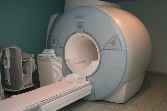
Machines:
- Siemens Magnetom Vida 3T
- Siemens Avanto 1,5T
- Philips Intera 1,5T
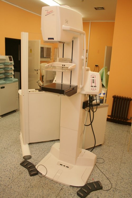
Breast mammography and sonography
Ordering patients (records):
Tel.: 224 438 145
Chief doctor
MD Daniel Zoubek
Tel.: 224 438 169
Email: daniel.zoubek@fnmotol.cz
At the workplace, diagnostic mammography is performed on a Planmed Sofie device and mammosonography on a Toshiba Nemio device.
Mammography performances:
- basic or additional projection mammography
- ductography
- stereotactically navigated preoperative localization of lesions with a wire
- stereotactically navigated core cut biopsies - classic 14G needle or vacuum biopsy with 10G needle
Ultrasound performances:
- basic examination or supplementary to mammography
- US guided preoperative localization - wires or radiopharmaceuticals (in cooperation with the Department of Nuclear Medicine 2nd Faculty of Medicine UK and FN Motol)
- US guided core cut biopsy and cyst puncture
Female patients come mainly from two mammo clinics and the oncology clinic at FNM, less from other FNM workplaces and also from extramural workplaces. Order times are item no. about 1 - 2 weeks for mammography and 1 - 2 months for ultrasound for non-acute examination. Cases requiring an earlier examination are carried out immediately, within a week at the latest.
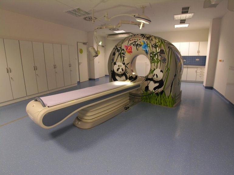
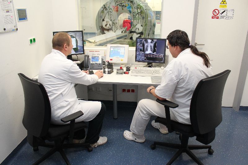
Pediatric computed tomography
Chief doctor
MD Jana Svítilová
tel .: (224) 438 174
Email: jana.svitilova@fnmotol.cz
Records (patient ordering)
tel .: (224) 435 072
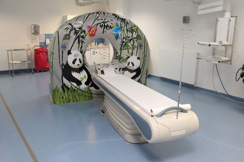
The premises of the Department of Pediatric Radiology are equipped with regard to pediatric patients. Na Homolce Hospital for verification and visualization of brain aneurysms.
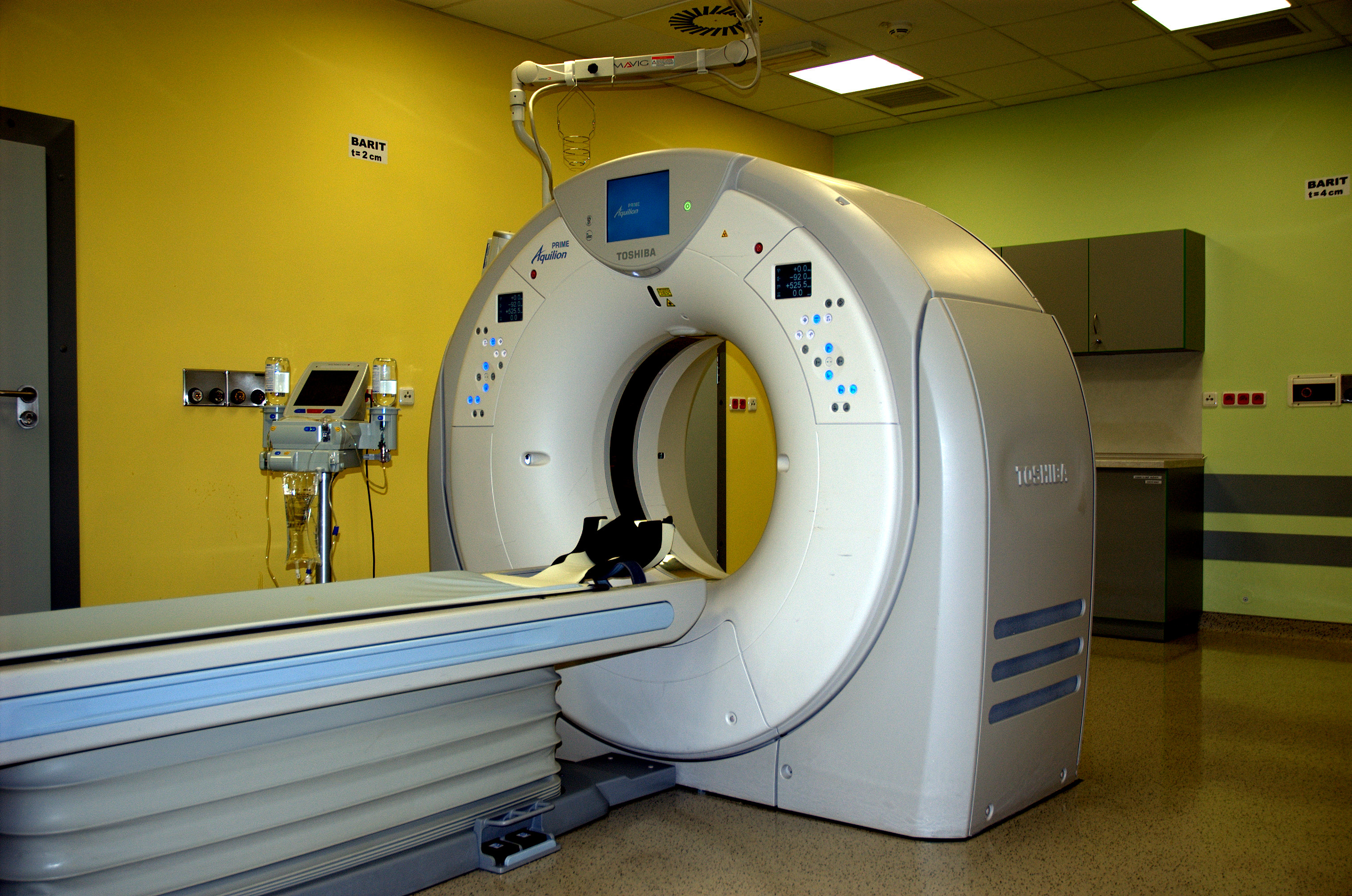
The Aquilion 64 computed tomograph (Toshiba) is installed at the workplace with technical parameters enabling high-quality, detailed, low-dose examinations to be performed on pediatric-friendly protocols.
In addition to interventional procedures under CT control, CT colonography and CT examination of the heart and intervention methods, the workplace performs routine and specialized examinations, including dental CT, incl. 3D visualization, virtual bronchoscopy, imaging of stimulation electrodes for subsequent MR fusion and CT angiography, including endovascular imaging.
The top Vitrea 130 workstation is used to evaluate the examination.
Examinations of pediatric patients are performed by erudite RAs and evaluated by physicians specializing in pediatric radiology.
Within the Department of Imaging Methods, the Department of Pediatric CT performs around 2500 examinations per year
Department of Pediatric Magnetic Resonance
Chief doctor
MUDr. Martin Kynčl Ph.D.
Email: martin.kyncl@fnmotol.cz
tel .: (224) 43 5030
Records (patient ordering)
tel .: (224) 43 5031
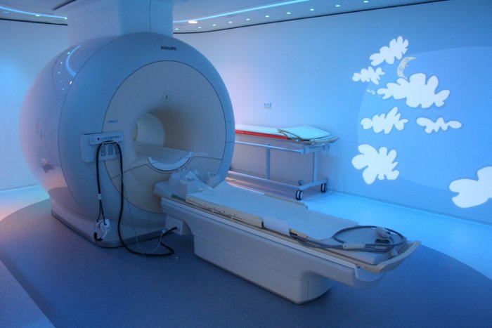
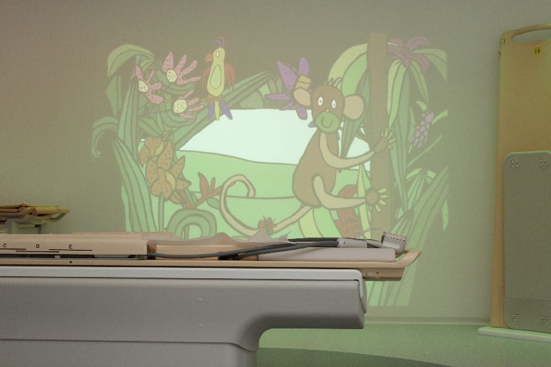
The Department of Pediatric Magnetic Resonance is now part of the pediatric department of the Clinic of Imaging Methods. The department is equipped with an Achieva 1.5T MR instrument from Philips using the most modern complementary techniques of color tuning of the examination room (so-called ambient lighting). The department is completely technically and personally equipped for examination and diagnostic imaging of pathologies in the youngest children, including examinations under general anesthesia.
The diagnostic spectrum includes complete pediatric oncology, neurology, pediatric and surgical issues in close cooperation with the clinical and research institutes of the University Hospital in Motol. A specialized team of doctors and laboratory technicians with high erudition in pediatric radiology also performs complicated examinations in premature babies, the department is part of the prenatal imaging center, it also performs spectroscopic, tractographic and functional MR examinations in children.
The Department of Pediatric Magnetic Resonance provides a diagnostic service for complicated examinations of childhood for Prague and the Central Bohemian Region, including examinations performed under general anesthesia.
The department performs a total of over 50 highly specialized pediatric MR examinations per week, of which at least 26 are under general anesthesia.
Operating time
Outpatient operation
Mon-Fri 7:00 - 15:00
Social activities
One of the main activities of the clinic is the undergraduate teaching of imaging methods for IV students. year 2. LF CU ended with an exam, preparation of students VI. year of the 2nd LF for the radiodiagnostic part of the state final exam in pediatrics and teaching to deepen the knowledge of 2nd LF students who chose imaging methods as an optional subject.
KZM doctors participate in postgraduate education in the basic field of Radiology and imaging methods, as well as in additional fields of Neuroradiology, Pediatric Radiology and Interventional Radiology.
Pedagogical clinics also often give lectures to students of the University of the 3rd Age. KZM employees actively participate in congresses, publish in foreign literature and organize or participate in the organization of Czech and foreign congresses.
The pediatric part of the clinic has a privileged position within the Czech Republic, whose staff methodically lead pediatric radiologists throughout the Czech Republic, provide teaching of pediatric radiology as part of postgraduate education and have a large share in the overall pre-certification preparation of Czech radiologists.
In cooperation with the International Atomic Energy Agency and the State Office for Nuclear Safety, the clinic regularly organizes internships for our and foreign experts training in the field of radiation protection.
Gallery
 Skiagraphy department - skiagraphic ...
 Skiagraphic workplace adult part –...
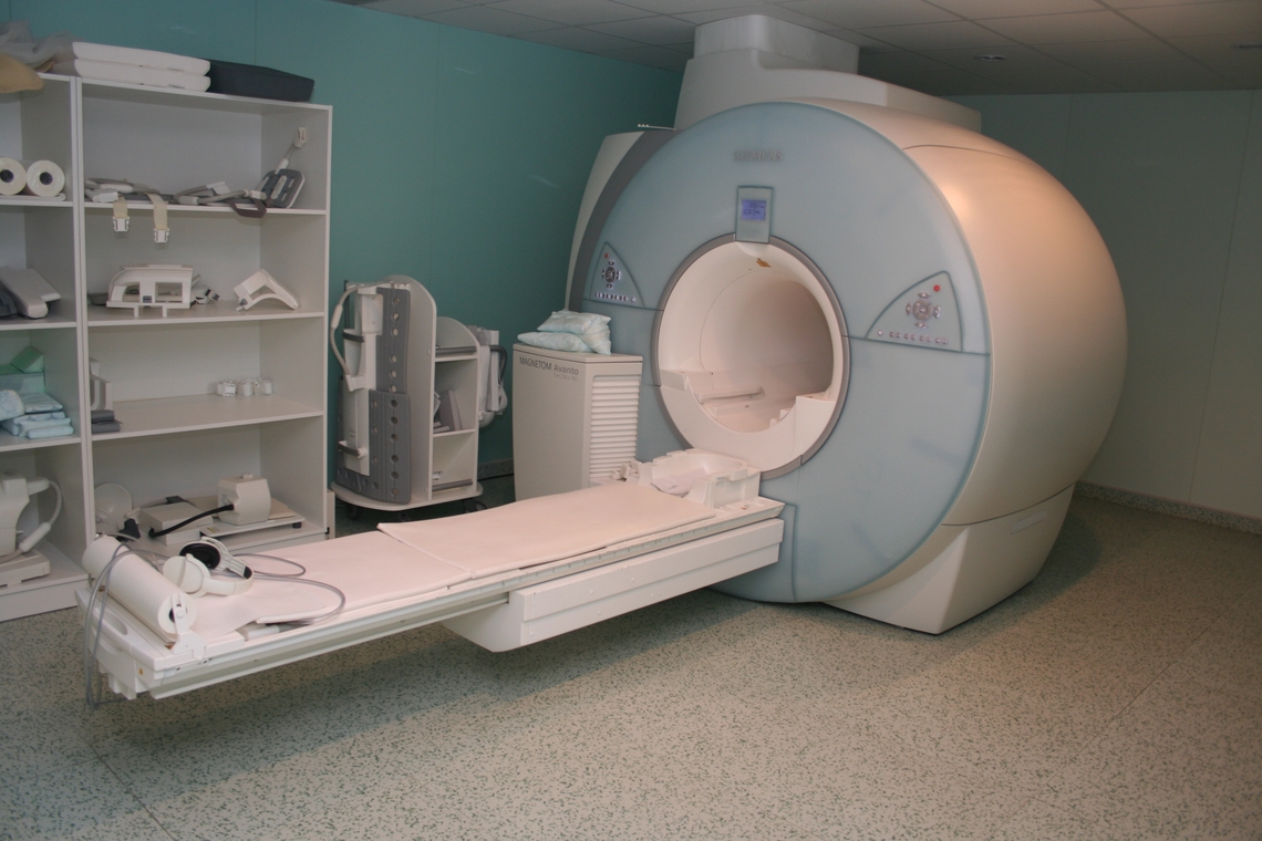 Department of Adult Magnetic Resonance ...
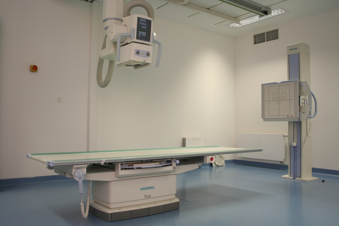 Skiagraphy department - skiagraphic ...
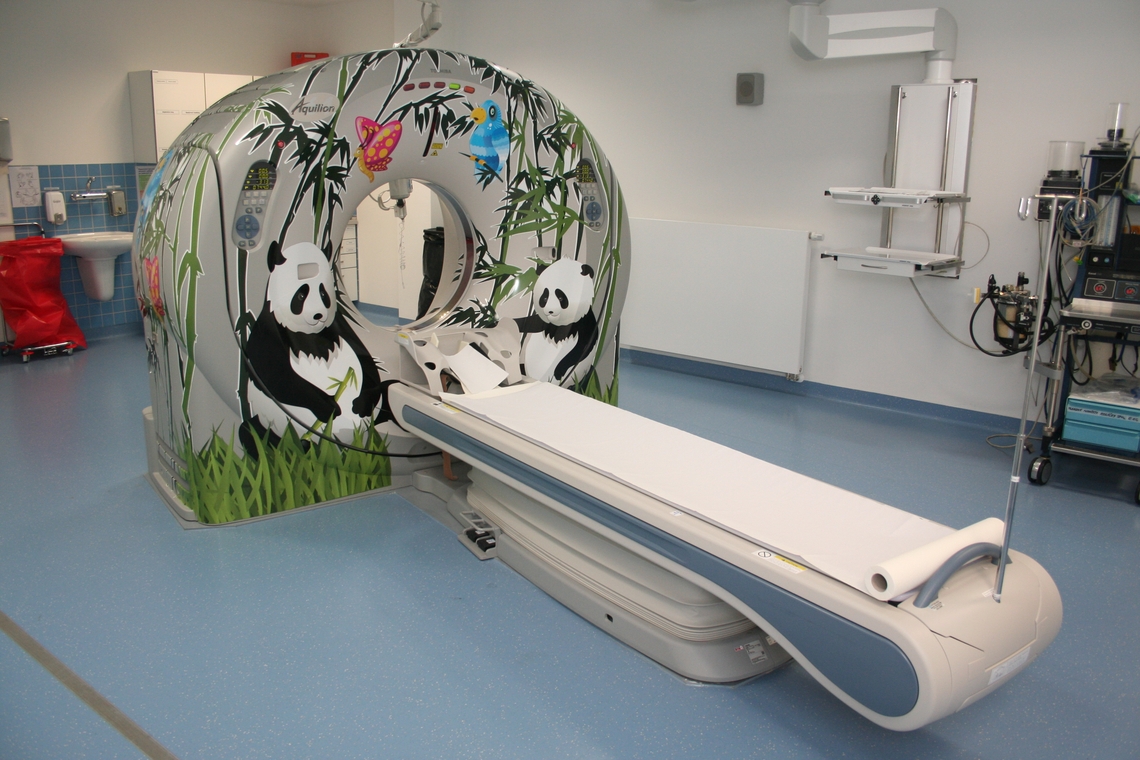 Department of Pediatric Computed Tomography ...
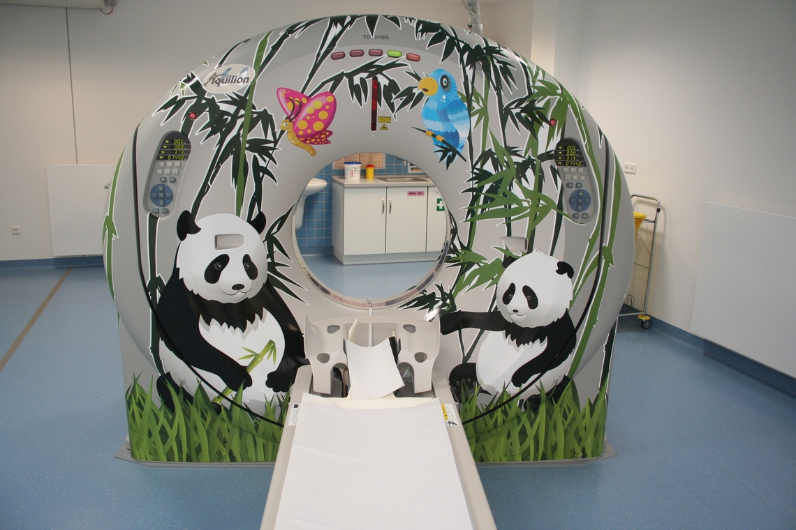 Department of Pediatric Computed Tomography ...
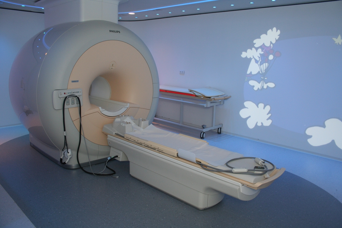 Department of Pediatric Magnetic Resonance ...
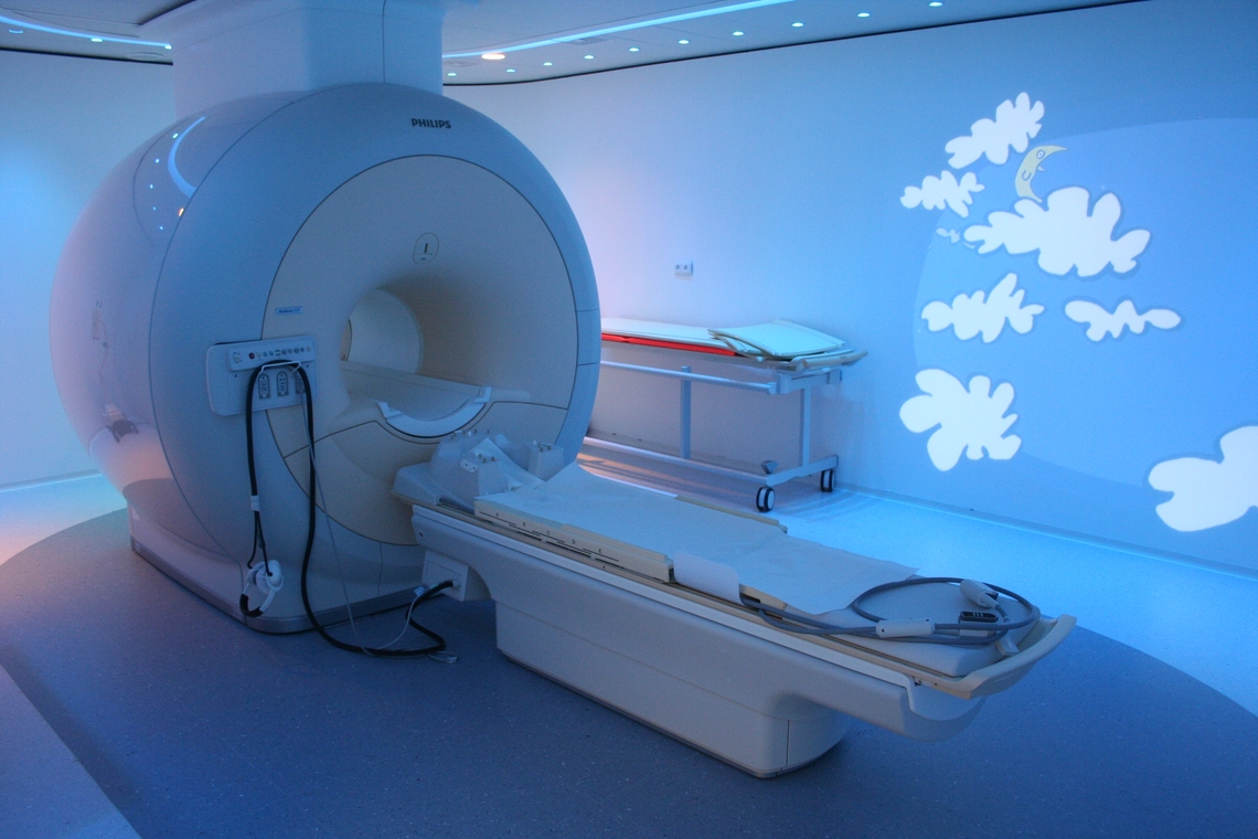 Department of Pediatric Magnetic Resonance ...
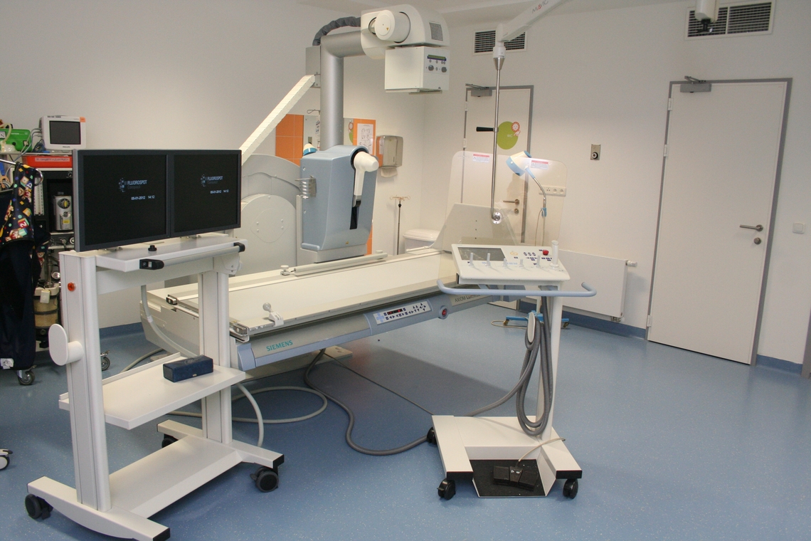 Department of Pediatric Conventional Radiology ...
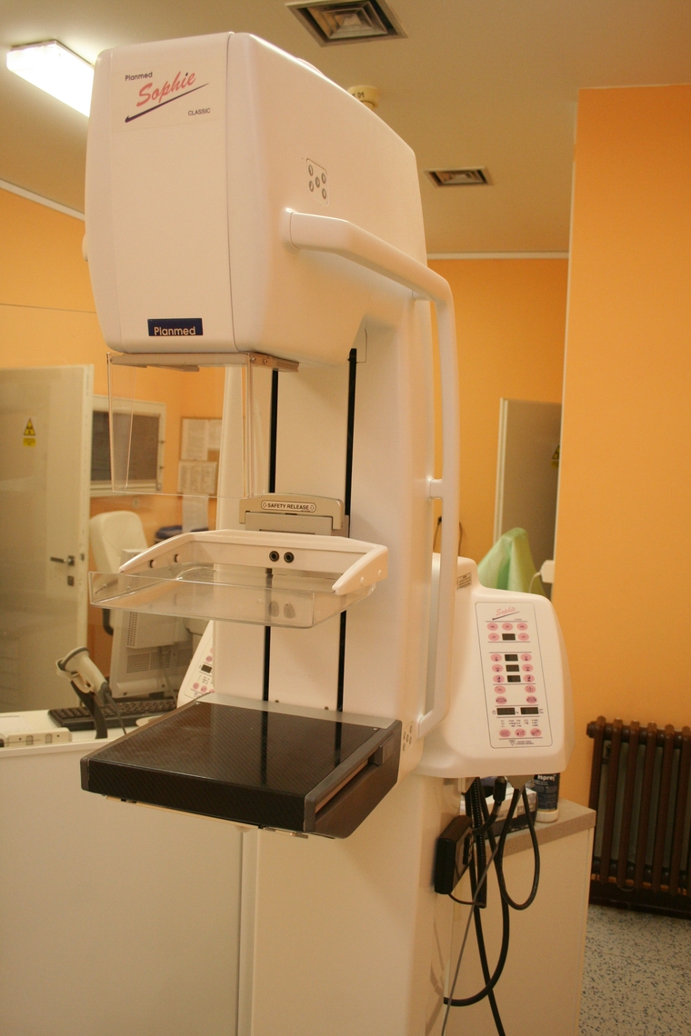 Mammography is performed at the workplace ...
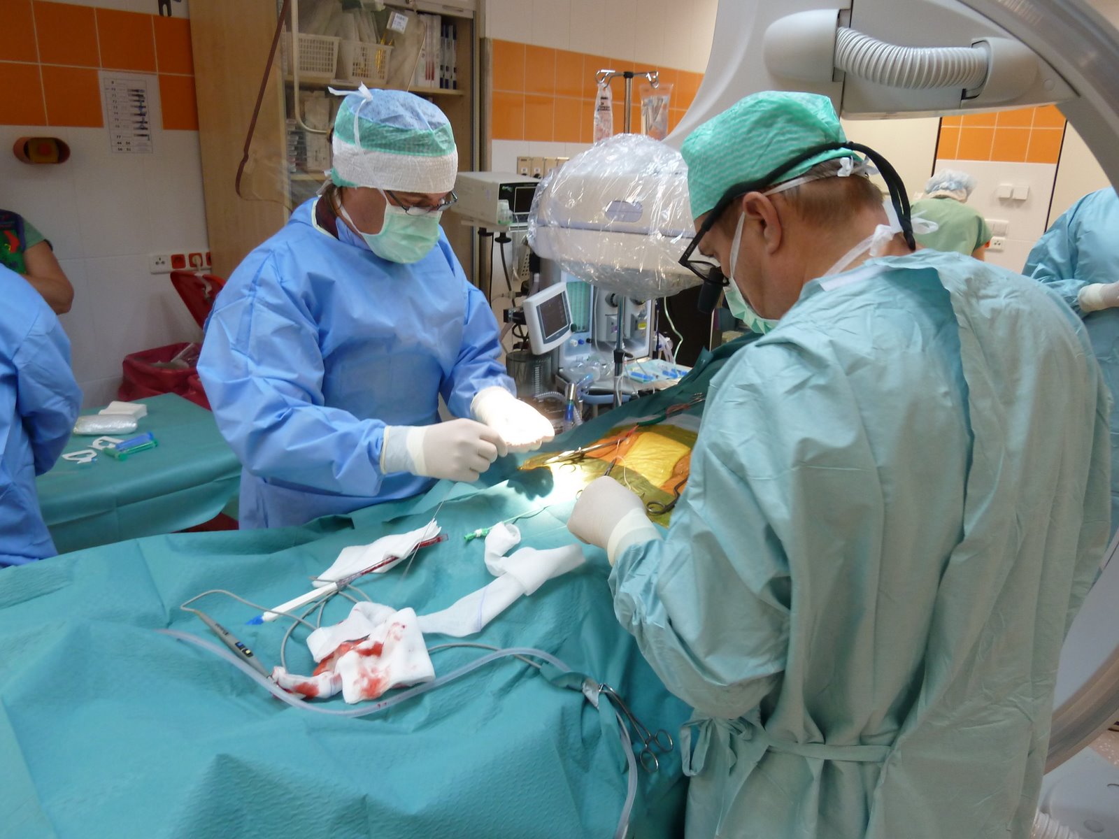 Department of Angiography - Hybrid Hall ....
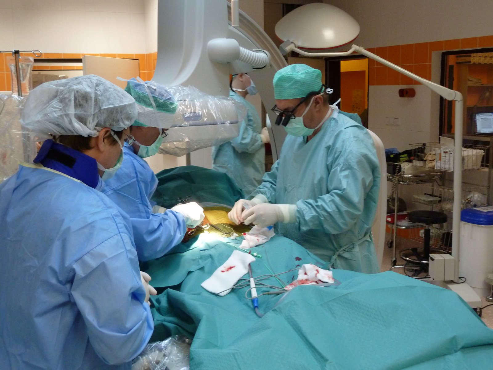 Angiography workplace, control room - view ...
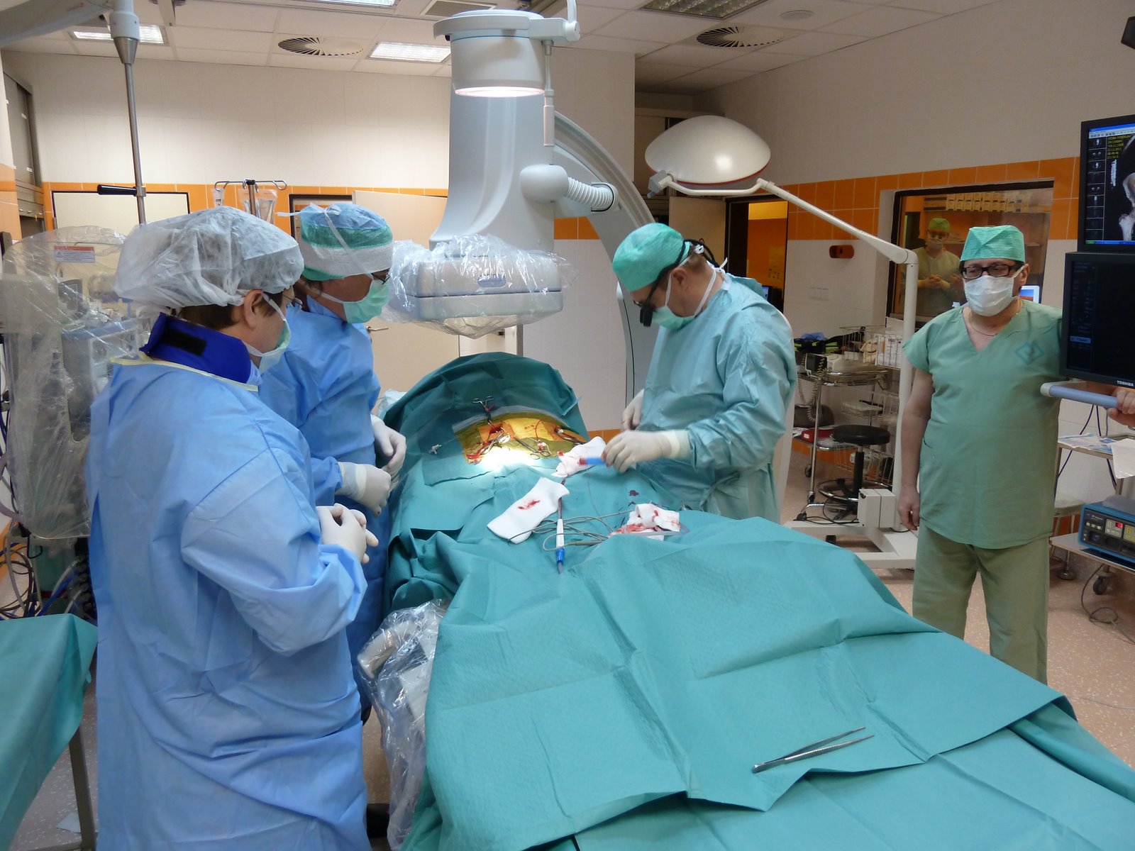 Department of Angiography - Hybrid Hall ....
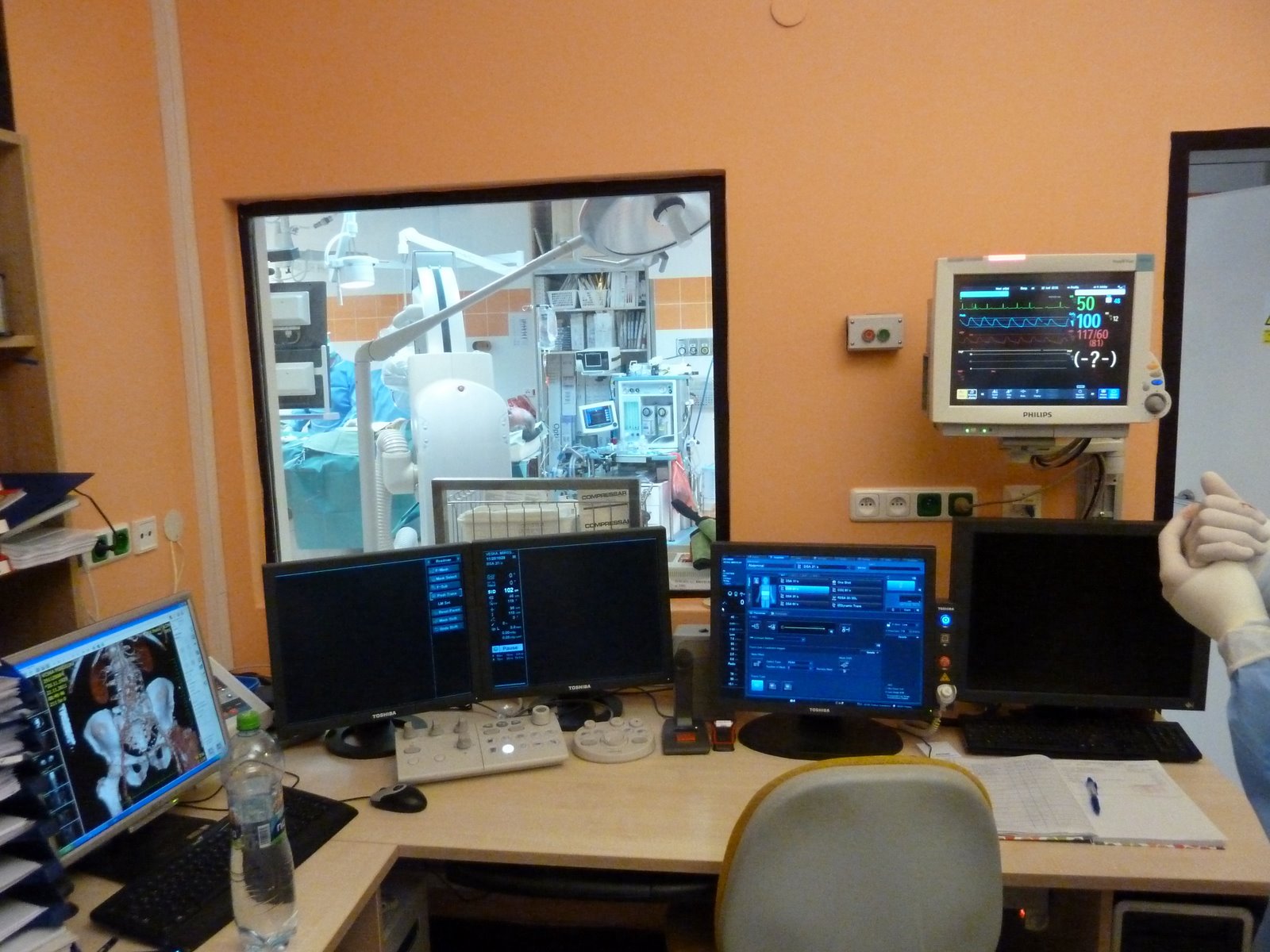 Department of Angiography - Hybrid Hall ....
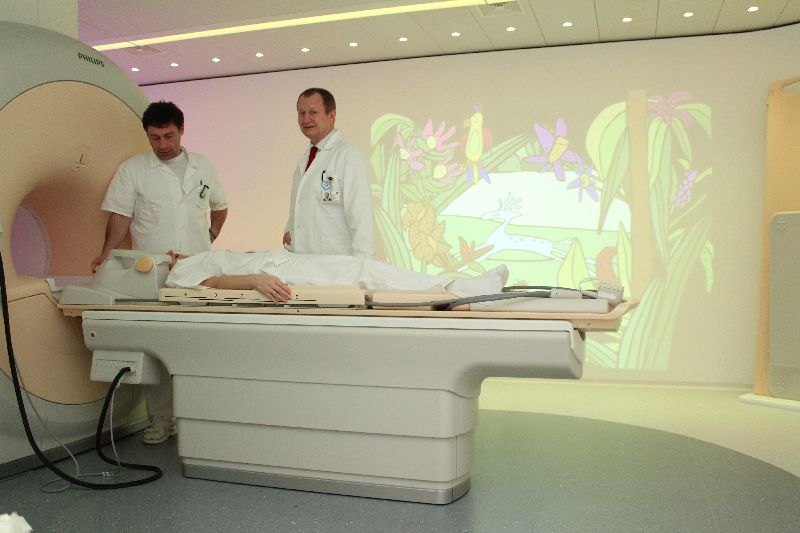
Contact
Email requests for sending images to ePACS to: epacs@fnmotol.cz
The head
prof. MD Miloslav Roček, CSc., MBA
Tel.: 224 438 100
Fax: 224 438 120
Email: miloslav.rocek@lfmotol.cuni.cz
Medical Deputy Chief for Adult
MUDr. Vojtech Suchanek
Tel.: 224 438 103
Email: vojtech.suchanek@fnmotol.cz
Medical deputy head for the children's section
MD Irena Buksakowska, Ph.D.
Tel.: 224 438 100, 224 438 101
Email: irena.buksakowska@fnmotol.cz
School deputy head of the clinic
MUDr. Vojtech Suchanek
Tel.: 224 438 103
Email: vojtech.suchanek@fnmotol.cz
Deputy head of the clinic for science and research
MD Martin Kynčl, Ph.D.
Tel.: 224 435 025
Email: martin.kyncl@fnmotol.cz
Senior radiology assistant - adult part
Mgr. Tomas Schilla
Tel.: 224 438 105
Email: tomas.schilla@fnmotol.cz
Senior radiology assistant - children's department
Alice Spring
Tel: 224 435 021
Email: alice.jara@fnmotol.cz
Clinic secretariat
Olga Skalicka, DiS.
Tel.: 224 438 100, 224 438 101
Fax.: 224 438 120
Evidence
Adult part
Tel.: 224 438 133, 224 438 124
Fax: 224 438 125
Children's part
Tel.: 224 435 012